
14.7: Polypeptides and Proteins
- Page ID
- 341985

\( \newcommand{\vecs}[1]{\overset { \scriptstyle \rightharpoonup} {\mathbf{#1}} } \)
\( \newcommand{\vecd}[1]{\overset{-\!-\!\rightharpoonup}{\vphantom{a}\smash {#1}}} \)
\( \newcommand{\dsum}{\displaystyle\sum\limits} \)
\( \newcommand{\dint}{\displaystyle\int\limits} \)
\( \newcommand{\dlim}{\displaystyle\lim\limits} \)
\( \newcommand{\id}{\mathrm{id}}\) \( \newcommand{\Span}{\mathrm{span}}\)
( \newcommand{\kernel}{\mathrm{null}\,}\) \( \newcommand{\range}{\mathrm{range}\,}\)
\( \newcommand{\RealPart}{\mathrm{Re}}\) \( \newcommand{\ImaginaryPart}{\mathrm{Im}}\)
\( \newcommand{\Argument}{\mathrm{Arg}}\) \( \newcommand{\norm}[1]{\| #1 \|}\)
\( \newcommand{\inner}[2]{\langle #1, #2 \rangle}\)
\( \newcommand{\Span}{\mathrm{span}}\)
\( \newcommand{\id}{\mathrm{id}}\)
\( \newcommand{\Span}{\mathrm{span}}\)
\( \newcommand{\kernel}{\mathrm{null}\,}\)
\( \newcommand{\range}{\mathrm{range}\,}\)
\( \newcommand{\RealPart}{\mathrm{Re}}\)
\( \newcommand{\ImaginaryPart}{\mathrm{Im}}\)
\( \newcommand{\Argument}{\mathrm{Arg}}\)
\( \newcommand{\norm}[1]{\| #1 \|}\)
\( \newcommand{\inner}[2]{\langle #1, #2 \rangle}\)
\( \newcommand{\Span}{\mathrm{span}}\) \( \newcommand{\AA}{\unicode[.8,0]{x212B}}\)
\( \newcommand{\vectorA}[1]{\vec{#1}} % arrow\)
\( \newcommand{\vectorAt}[1]{\vec{\text{#1}}} % arrow\)
\( \newcommand{\vectorB}[1]{\overset { \scriptstyle \rightharpoonup} {\mathbf{#1}} } \)
\( \newcommand{\vectorC}[1]{\textbf{#1}} \)
\( \newcommand{\vectorD}[1]{\overrightarrow{#1}} \)
\( \newcommand{\vectorDt}[1]{\overrightarrow{\text{#1}}} \)
\( \newcommand{\vectE}[1]{\overset{-\!-\!\rightharpoonup}{\vphantom{a}\smash{\mathbf {#1}}}} \)
\( \newcommand{\vecs}[1]{\overset { \scriptstyle \rightharpoonup} {\mathbf{#1}} } \)
\(\newcommand{\longvect}{\overrightarrow}\)
\( \newcommand{\vecd}[1]{\overset{-\!-\!\rightharpoonup}{\vphantom{a}\smash {#1}}} \)
\(\newcommand{\avec}{\mathbf a}\) \(\newcommand{\bvec}{\mathbf b}\) \(\newcommand{\cvec}{\mathbf c}\) \(\newcommand{\dvec}{\mathbf d}\) \(\newcommand{\dtil}{\widetilde{\mathbf d}}\) \(\newcommand{\evec}{\mathbf e}\) \(\newcommand{\fvec}{\mathbf f}\) \(\newcommand{\nvec}{\mathbf n}\) \(\newcommand{\pvec}{\mathbf p}\) \(\newcommand{\qvec}{\mathbf q}\) \(\newcommand{\svec}{\mathbf s}\) \(\newcommand{\tvec}{\mathbf t}\) \(\newcommand{\uvec}{\mathbf u}\) \(\newcommand{\vvec}{\mathbf v}\) \(\newcommand{\wvec}{\mathbf w}\) \(\newcommand{\xvec}{\mathbf x}\) \(\newcommand{\yvec}{\mathbf y}\) \(\newcommand{\zvec}{\mathbf z}\) \(\newcommand{\rvec}{\mathbf r}\) \(\newcommand{\mvec}{\mathbf m}\) \(\newcommand{\zerovec}{\mathbf 0}\) \(\newcommand{\onevec}{\mathbf 1}\) \(\newcommand{\real}{\mathbb R}\) \(\newcommand{\twovec}[2]{\left[\begin{array}{r}#1 \\ #2 \end{array}\right]}\) \(\newcommand{\ctwovec}[2]{\left[\begin{array}{c}#1 \\ #2 \end{array}\right]}\) \(\newcommand{\threevec}[3]{\left[\begin{array}{r}#1 \\ #2 \\ #3 \end{array}\right]}\) \(\newcommand{\cthreevec}[3]{\left[\begin{array}{c}#1 \\ #2 \\ #3 \end{array}\right]}\) \(\newcommand{\fourvec}[4]{\left[\begin{array}{r}#1 \\ #2 \\ #3 \\ #4 \end{array}\right]}\) \(\newcommand{\cfourvec}[4]{\left[\begin{array}{c}#1 \\ #2 \\ #3 \\ #4 \end{array}\right]}\) \(\newcommand{\fivevec}[5]{\left[\begin{array}{r}#1 \\ #2 \\ #3 \\ #4 \\ #5 \\ \end{array}\right]}\) \(\newcommand{\cfivevec}[5]{\left[\begin{array}{c}#1 \\ #2 \\ #3 \\ #4 \\ #5 \\ \end{array}\right]}\) \(\newcommand{\mattwo}[4]{\left[\begin{array}{rr}#1 \amp #2 \\ #3 \amp #4 \\ \end{array}\right]}\) \(\newcommand{\laspan}[1]{\text{Span}\{#1\}}\) \(\newcommand{\bcal}{\cal B}\) \(\newcommand{\ccal}{\cal C}\) \(\newcommand{\scal}{\cal S}\) \(\newcommand{\wcal}{\cal W}\) \(\newcommand{\ecal}{\cal E}\) \(\newcommand{\coords}[2]{\left\{#1\right\}_{#2}}\) \(\newcommand{\gray}[1]{\color{gray}{#1}}\) \(\newcommand{\lgray}[1]{\color{lightgray}{#1}}\) \(\newcommand{\rank}{\operatorname{rank}}\) \(\newcommand{\row}{\text{Row}}\) \(\newcommand{\col}{\text{Col}}\) \(\renewcommand{\row}{\text{Row}}\) \(\newcommand{\nul}{\text{Nul}}\) \(\newcommand{\var}{\text{Var}}\) \(\newcommand{\corr}{\text{corr}}\) \(\newcommand{\len}[1]{\left|#1\right|}\) \(\newcommand{\bbar}{\overline{\bvec}}\) \(\newcommand{\bhat}{\widehat{\bvec}}\) \(\newcommand{\bperp}{\bvec^\perp}\) \(\newcommand{\xhat}{\widehat{\xvec}}\) \(\newcommand{\vhat}{\widehat{\vvec}}\) \(\newcommand{\uhat}{\widehat{\uvec}}\) \(\newcommand{\what}{\widehat{\wvec}}\) \(\newcommand{\Sighat}{\widehat{\Sigma}}\) \(\newcommand{\lt}{<}\) \(\newcommand{\gt}{>}\) \(\newcommand{\amp}{&}\) \(\definecolor{fillinmathshade}{gray}{0.9}\)- Define or describe the following:
- amino acid
- "R" group
- peptide bond
- peptide
- polypeptide
- primary protein structure
- secondary protein structure
- tertiary protein structure
- quaternary protein structure
- gene
- Describe how the primary structure of a protein or polypeptide ultimately detemines its final three-dimensional shape.
- Describe how the order of nucleotide bases in DNA ultimately determines the final three-dimensional shape of a protein or polypeptide.
Amino acids are the building blocks for proteins. All amino acids contain an amino or NH2 group and a carboxyl (acid) or COOH group. There are 20 different amino acids commonly found in proteins and often 300 or more amino acids per protein molecule. Each amino acid differs in terms of its "R" group. The "R" group of an amino acid is the remainder of the molecule, that is, the portion other than the amino group, the acid group, and the central carbon. Each different amino acid has a unique "R" group and the unique chemical properties of an amino acid depend on that of its "R" group (Figure \(\PageIndex{1}\)).
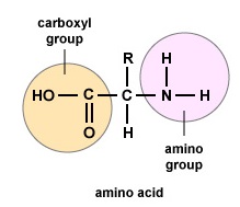 Figure \(\PageIndex{1}\): Amino Acids. Structure of an amino acid.
To form polypeptides and proteins, amino acids are joined together by peptide bonds, in which the amino or NH2 of one amino acid bonds to the carboxyl (acid) or COOH group of another amino acid as shown in (Figure \(\PageIndex{2}\) and Figure \(\PageIndex{3}\)).
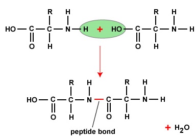 Figure \(\PageIndex{2}\): Peptide Bonds. A peptide bond forms when the amino group of one amino acid bonds to the carboxyl group of another amino acid.
A peptide is two or more amino acids joined together by peptide bonds, and a polypeptide is a chain of many amino acids. A protein contains one or more polypeptides. Therefore, proteins are long chains of amino acids held together by peptide bonds.
 Figure \(\PageIndex{3}\): Formation of a Peptide Bond. A peptide bond forms when the amino group of one amino acid bonds to the carboxyl group of another amino acid.
The actual order of the amino acids in the protein is called its primary structure (Figure \(\PageIndex{4}\)) and is determined by DNA. As will be seen later in this unit, DNA is divided into functional units called genes. A gene is a sequence of deoxyribonucleotide bases along one strand of DNA that codes for a functional product - a specific molecule of messenger RNA, transfer RNA, or ribosomal RNA. The product is usually messenger RNA (mRNA) and mRNA ultimately results in the synthesis of a polypeptide or a protein. Therefore, it is commonly said that the order of deoxyribonucleotide bases in a gene determines the amino acid sequence of a particular protein. Since certain amino acids can interact with other amino acids in the same protein, this primary structure ultimately determines the final shape and therefore the chemical and physical properties of the protein.
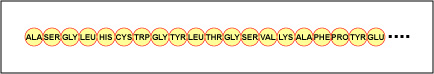 Figure \(\PageIndex{4}\): Primary Structure of a Protein or Polypeptide. The primary structure of a protein or polypeptide is the actual sequence of its amino acids. Primary structure is determined by the order of the deoxyribonucleotide bases in genes.
The secondary structure of the protein is due to hydrogen bonds that form between the oxygen atom of one amino acid and the nitrogen atom of another. This gives the protein or polypeptide the two-dimensional form of an alpha-helix or a beta-pleated sheet (Figure \(\PageIndex{5}\)).
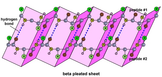 Figure \(\PageIndex{5}\): Secondary Structure of a Protein or Polypeptide. (left) The secondary structure of a protein or polypeptide is due to hydrogen bonds forming between an oxygen atom of one amino acid and a nitrogen atom of another. There are two possible types of secondary structure: an alpha helix and a beta sheet. In the case of an alpha helix, the hydrogen bonding causes the polypeptide to twist into a helix. With a beta sheet the hydrogen bonding enables the polypeptide to fold back and forth upon itself like a pleated sheet. (right) The secondary structure of a protein or polypeptide is due to hydrogen bonds forming between an oxygen atom of one amino acid and a nitrogen atom of another. There are two possible types of secondary structure: an alpha helix and a beta sheet. In the case of an alpha helix, the hydrogen bonding causes the polypeptide to twist into a helix. With a beta sheet the hydrogen bonding enables the polypeptide to fold back and forth upon itself like a pleated sheet.
In globular proteins such as enzymes, the long chain of amino acids becomes folded into a three-dimensional functional shape or tertiary structure. This is because certain amino acids with sulfhydryl or SH groups form disulfide (S-S) bonds with other amino acids in the same chain. Other interactions between R groups of amino acids such as hydrogen bonds, ionic bonds, covalent bonds, and hydrophobic interactions also contribute to the tertiary structure (Figure \(\PageIndex{6}\)). In some proteins, such as antibody molecules and hemoglobin, several polypeptides may bond together to form a quaternary structure (Figure \(\PageIndex{7}\)).
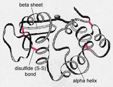 Figure \(\PageIndex{6}\): Tertiary Structure of a Protein or Polypeptide. In globular proteins such as enzymes, the long chain of amino acids becomes folded into a three-dimensional functional shape or tertiary structure. This is because certain amino acids with sulfhydryl or SH groups form disulfide (S-S) bonds with other amino acids in the same chain. Other interactions between R groups of amino acids such as hydrogen bonds, ionic bonds, covalent bonds, and hydrophobic interactions also contribute to the tertiary structure.
As will be seen later in this unit, during protein synthesis, the order of nucleotide bases along a gene gets transcribed into a complementary strand of mRNA which is then translated by tRNA into the correct order of amino acids for that polypeptide or protein. Therefore, the order of deoxyribonucleotide bases along the DNA determines the order of amino acids in the proteins. Because certain amino acids can interact with other amino acids, the order of amino acids for each protein determines its final three-dimensional shape, which in turn determines the function of that protein (e.g., what substrate an enzyme will react with, what epitopes the Fab of an antibody will combine with, what receptors a cytokine will bind to).
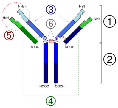 Figure \(\PageIndex{7}\): Quaternary Structure of a Protein. The quaternary structure of a protein is due to several polypeptides joining together, as in the case of antibody molecules. Schematic diagram of the basic unit of immunoglobulin (antibody) Fab Fc heavy chain (consist of VH, CH1, hinge, CH2 and CH3 regions: from N-term) light chain (consist of VL and CL regions: from N-term) antigen binding site hinge regions (*) -S-S- mean disulfide bonds. (CC-SA-BY 3.0; Y_tambe).
Summary
- Amino acids are the building blocks for proteins. There are 20 different amino acids commonly found in proteins and often 300 or more amino acids per protein molecule.
- All amino acids contain an amino or NH2 group and a carboxyl (acid) or COOH group.
- To form polypeptides and proteins, amino acids are joined together by peptide bonds, in which the amino or NH2 of one amino acid bonds to the carboxyl (acid) or COOH group of another amino acid.
- A peptide is two or more amino acids joined together by peptide bonds; a polypeptide is a chain of many amino acids; and a protein contains one or more polypeptides. Therefore, proteins are long chains of amino acids held together by peptide bonds.
- The actual order of the amino acids in the protein is called its primary structure and is determined by DNA.
- The order of deoxyribonucleotide bases in a gene determines the amino acid sequence of a particular protein. Since certain amino acids can interact with other amino acids in the same protein, this primary structure ultimately determines the final shape and therefore the chemical and physical properties of the protein.
- The secondary structure of the protein is due to hydrogen bonds that form between the oxygen atom of one amino acid and the nitrogen atom of another and gives the protein or polypeptide the two-dimensional form of an alpha-helix or a beta-pleated sheet.
- In globular proteins such as enzymes, the long chain of amino acids becomes folded into a three-dimensional functional shape or tertiary structure. This is because certain amino acids with sulfhydryl or SH groups form disulfide (S-S) bonds with other amino acids in the same chain. Other interactions between R groups of amino acids such as hydrogen bonds, ionic bonds, covalent bonds, and hydrophobic interactions also contribute to the tertiary structure.
- In some proteins, such as antibody molecules, several polypeptides may bond together to form a quaternary structure.