
15.5.2: Transport in Membranes
- Page ID
- 306712

\( \newcommand{\vecs}[1]{\overset { \scriptstyle \rightharpoonup} {\mathbf{#1}} } \)
\( \newcommand{\vecd}[1]{\overset{-\!-\!\rightharpoonup}{\vphantom{a}\smash {#1}}} \)
\( \newcommand{\id}{\mathrm{id}}\) \( \newcommand{\Span}{\mathrm{span}}\)
( \newcommand{\kernel}{\mathrm{null}\,}\) \( \newcommand{\range}{\mathrm{range}\,}\)
\( \newcommand{\RealPart}{\mathrm{Re}}\) \( \newcommand{\ImaginaryPart}{\mathrm{Im}}\)
\( \newcommand{\Argument}{\mathrm{Arg}}\) \( \newcommand{\norm}[1]{\| #1 \|}\)
\( \newcommand{\inner}[2]{\langle #1, #2 \rangle}\)
\( \newcommand{\Span}{\mathrm{span}}\)
\( \newcommand{\id}{\mathrm{id}}\)
\( \newcommand{\Span}{\mathrm{span}}\)
\( \newcommand{\kernel}{\mathrm{null}\,}\)
\( \newcommand{\range}{\mathrm{range}\,}\)
\( \newcommand{\RealPart}{\mathrm{Re}}\)
\( \newcommand{\ImaginaryPart}{\mathrm{Im}}\)
\( \newcommand{\Argument}{\mathrm{Arg}}\)
\( \newcommand{\norm}[1]{\| #1 \|}\)
\( \newcommand{\inner}[2]{\langle #1, #2 \rangle}\)
\( \newcommand{\Span}{\mathrm{span}}\) \( \newcommand{\AA}{\unicode[.8,0]{x212B}}\)
\( \newcommand{\vectorA}[1]{\vec{#1}} % arrow\)
\( \newcommand{\vectorAt}[1]{\vec{\text{#1}}} % arrow\)
\( \newcommand{\vectorB}[1]{\overset { \scriptstyle \rightharpoonup} {\mathbf{#1}} } \)
\( \newcommand{\vectorC}[1]{\textbf{#1}} \)
\( \newcommand{\vectorD}[1]{\overrightarrow{#1}} \)
\( \newcommand{\vectorDt}[1]{\overrightarrow{\text{#1}}} \)
\( \newcommand{\vectE}[1]{\overset{-\!-\!\rightharpoonup}{\vphantom{a}\smash{\mathbf {#1}}}} \)
\( \newcommand{\vecs}[1]{\overset { \scriptstyle \rightharpoonup} {\mathbf{#1}} } \)
\( \newcommand{\vecd}[1]{\overset{-\!-\!\rightharpoonup}{\vphantom{a}\smash {#1}}} \)
\(\newcommand{\avec}{\mathbf a}\) \(\newcommand{\bvec}{\mathbf b}\) \(\newcommand{\cvec}{\mathbf c}\) \(\newcommand{\dvec}{\mathbf d}\) \(\newcommand{\dtil}{\widetilde{\mathbf d}}\) \(\newcommand{\evec}{\mathbf e}\) \(\newcommand{\fvec}{\mathbf f}\) \(\newcommand{\nvec}{\mathbf n}\) \(\newcommand{\pvec}{\mathbf p}\) \(\newcommand{\qvec}{\mathbf q}\) \(\newcommand{\svec}{\mathbf s}\) \(\newcommand{\tvec}{\mathbf t}\) \(\newcommand{\uvec}{\mathbf u}\) \(\newcommand{\vvec}{\mathbf v}\) \(\newcommand{\wvec}{\mathbf w}\) \(\newcommand{\xvec}{\mathbf x}\) \(\newcommand{\yvec}{\mathbf y}\) \(\newcommand{\zvec}{\mathbf z}\) \(\newcommand{\rvec}{\mathbf r}\) \(\newcommand{\mvec}{\mathbf m}\) \(\newcommand{\zerovec}{\mathbf 0}\) \(\newcommand{\onevec}{\mathbf 1}\) \(\newcommand{\real}{\mathbb R}\) \(\newcommand{\twovec}[2]{\left[\begin{array}{r}#1 \\ #2 \end{array}\right]}\) \(\newcommand{\ctwovec}[2]{\left[\begin{array}{c}#1 \\ #2 \end{array}\right]}\) \(\newcommand{\threevec}[3]{\left[\begin{array}{r}#1 \\ #2 \\ #3 \end{array}\right]}\) \(\newcommand{\cthreevec}[3]{\left[\begin{array}{c}#1 \\ #2 \\ #3 \end{array}\right]}\) \(\newcommand{\fourvec}[4]{\left[\begin{array}{r}#1 \\ #2 \\ #3 \\ #4 \end{array}\right]}\) \(\newcommand{\cfourvec}[4]{\left[\begin{array}{c}#1 \\ #2 \\ #3 \\ #4 \end{array}\right]}\) \(\newcommand{\fivevec}[5]{\left[\begin{array}{r}#1 \\ #2 \\ #3 \\ #4 \\ #5 \\ \end{array}\right]}\) \(\newcommand{\cfivevec}[5]{\left[\begin{array}{c}#1 \\ #2 \\ #3 \\ #4 \\ #5 \\ \end{array}\right]}\) \(\newcommand{\mattwo}[4]{\left[\begin{array}{rr}#1 \amp #2 \\ #3 \amp #4 \\ \end{array}\right]}\) \(\newcommand{\laspan}[1]{\text{Span}\{#1\}}\) \(\newcommand{\bcal}{\cal B}\) \(\newcommand{\ccal}{\cal C}\) \(\newcommand{\scal}{\cal S}\) \(\newcommand{\wcal}{\cal W}\) \(\newcommand{\ecal}{\cal E}\) \(\newcommand{\coords}[2]{\left\{#1\right\}_{#2}}\) \(\newcommand{\gray}[1]{\color{gray}{#1}}\) \(\newcommand{\lgray}[1]{\color{lightgray}{#1}}\) \(\newcommand{\rank}{\operatorname{rank}}\) \(\newcommand{\row}{\text{Row}}\) \(\newcommand{\col}{\text{Col}}\) \(\renewcommand{\row}{\text{Row}}\) \(\newcommand{\nul}{\text{Nul}}\) \(\newcommand{\var}{\text{Var}}\) \(\newcommand{\corr}{\text{corr}}\) \(\newcommand{\len}[1]{\left|#1\right|}\) \(\newcommand{\bbar}{\overline{\bvec}}\) \(\newcommand{\bhat}{\widehat{\bvec}}\) \(\newcommand{\bperp}{\bvec^\perp}\) \(\newcommand{\xhat}{\widehat{\xvec}}\) \(\newcommand{\vhat}{\widehat{\vvec}}\) \(\newcommand{\uhat}{\widehat{\uvec}}\) \(\newcommand{\what}{\widehat{\wvec}}\) \(\newcommand{\Sighat}{\widehat{\Sigma}}\) \(\newcommand{\lt}{<}\) \(\newcommand{\gt}{>}\) \(\newcommand{\amp}{&}\) \(\definecolor{fillinmathshade}{gray}{0.9}\)Source: BiochemFFA_3_2.pdf. The entire textbook is available for free from the authors at http://biochem.science.oregonstate.edu/content/biochemistry-free-and-easy
Movement of materials across membranes
As noted earlier, it is essential for cells to be able to uptake nutrients. This function along with movement of ions and other substances is provided by proteins/protein complexes that are highly specific for the compounds they move.
Selective movement of ions by membrane proteins and the ions’ extremely low permeability across the lipid bilayer are important for helping to maintain the osmotic balance of the cell and also for providing for the most important mechanism for it to make ATP - the process of oxidative phosphorylation.
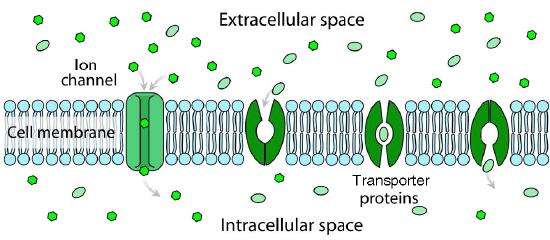
Channels and transporters
With respect to movement of materials through membrane proteins, there is a difference between channels (sometimes called pores) and transporters. Channels largely provide openings with some specificity and molecules pass through them at close to the rate of diffusion. They usually involve movement of water or ions. Examples would be the sodium or potassium channels of nerve cells. Transporters have high specificity and transfer rates that are orders of magnitude slower. Transport proteins include the sodium-potassium pump, the sodium-calcium exchanger, and lactose permease, amongst many others).
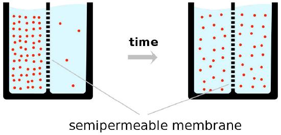
Facilitated diffusion
As noted, the driving force for facilitated diffusion is concentration, meaning that in facilitated diffusion, materials will only move from a higher concentration to a lower concentration and that at the end of the process, the concentration of materials on each side of a bilayer will be equal (Figure 3.28). This may work well in many cases.
For example, the blood concentration of glucose is sufficiently high that red blood cells can use facilitated diffusion as a means of acquiring glucose. Other cells, further removed from the blood supply where the glucose concentration is lower, must use active transport mechanisms because there is not a sufficient concentration of glucose to provide cells with the glucose they need.
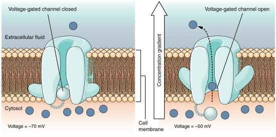
Ion channels
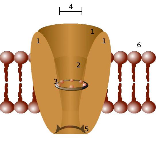
Ion channels are pore-forming membrane proteins in the membranes of all cells that regulate movement of selected ions across a membrane (Figures 3.29 & 3.30). They help to establish the resting membrane potential and to affect action potentials and other electrical signals. They are very important in the process of nerve transmission. Ion channels control the flow of ions across secretory and epithelial cells, and consequently help to regulate cell volume by affecting osmotic pressure.
Ion channels are essential features of almost all cells, functioning as selective “tunnels” that restrict movement through them to ions with specific characteristics (typically size). The size of the opening is very narrow (usually one or two atoms wide) and is able to select even against ions that are too small.
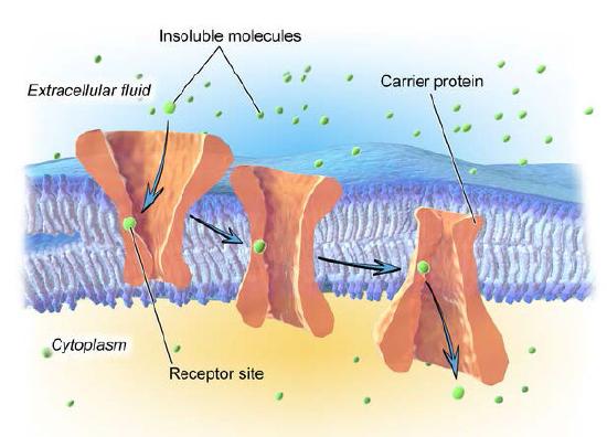
Transporter proteins
Not all facilitated transport occurs through ion channel proteins. Transporter proteins, as noted earlier (HERE and Figure 3.27) facilitate movement of materials across a lipid bilayer, but are slower than ion channels. Figure 3.36 illustrates a transporter protein in action. As can be seen, transporter proteins rely on a specific receptor site for proper recognition of the molecule to be moved.
Binding of the proper molecule causes a conformational change in the shape of the protein (an eversion) which results in a flipping of the open side of the protein to the other side of the lipid bilayer. In this way, the molecule is moved. Like ion channels, transporter proteins facilitate movement of materials in either direction, driven only by the concentration difference between one side and the other.
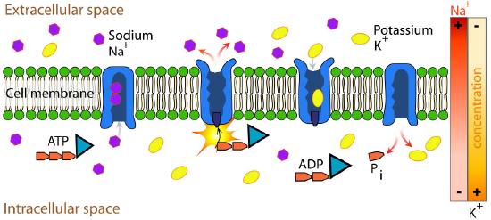
Active transport
All of the transport mechanisms described so far are driven solely by a concentration gradient - moving from higher concentrations in the direction of lower concentrations. These movements can occur in either direction and, as noted, result in equal concentrations on either side of the bilayer, if allowed to go to completion. Many times, however, cells must move materials against a concentration gradient and when this occurs, another source of energy is required. This process is known as active transport.
A good definition of active transport is that in active transport, at least one molecule is being moved against a concentration gradient. A common, but not exclusive, energy source is ATP.
Nerve transmission
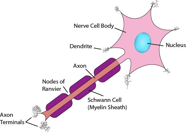
Now that you have seen how the Na+/K+ ATPase functions, it is appropriate to discuss how nerve cells use ion gradients created with it to generate and transmit nerve signals. Neurons are cells of the nervous system that use chemical and electrical signals to rapidly transmit information across the body (Figure 3.40). The sensory nerve system links receptors for vision, hearing, touch, taste, and smell to the brain for perception. Motor neurons run from the spinal cord to muscle cells. These neurons have a cell body and a very long, thin extension called an axon, that stretches from the cell body in the spinal cord all the way to the muscles they control. Nerve impulses travel down the axon to stimulate muscle contraction.
Signals travel through neurons, ultimately arriving at junctions with other nerve cells or target cells such as muscle cells. Note that neurons do not make physical contact with each other or with muscle cells. The tiny space between two neurons or between a neuron and a muscle cell is called the synaptic cleft. At the synaptic cleft, the neuron releases neurotransmitters that exit the nerve cell and travel across the junction to a recipient cell where a response is generated. That response may be creating another nerve signal, if the adjacent cell is a nerve cell or it may be a muscular contraction if the recipient is a muscle cell (Figure 3.41).
In considering information movement via nerve cells, then, we will discuss two steps - 1) creation and propagation of a signal in a nerve cell and 2) action of neurotransmitters exiting a nerve cell and transiting a synaptic junction.
Signal source
Creation of a nerve signal begins with a stimulus to the nerve cell. In the case of muscle contraction, the motor cortex of the brain sends signals to the appropriate motor neurons, stimulating them to generate a nerve impulse. How is such an impulse generated?
Resting potential
In the unstimulated state, all cells, including nerve cells, have a small voltage difference (called the resting potential) across the plasma membrane, arising from unequal pumping of ions across the membrane. The Na+/K+ ATPase, for example, pumps sodium ions out of the cell and potassium ions into cells. Since three sodium ions get pumped out for every two potassium ions pumped in, a charge and chemical gradient is created. It is the charge gradient that gives rise to the resting potential.
Altering the gradients of ions across membranes provide the driving force for nerve signals. This happens as a result of opening and closing of gated ion channels. Opening of gates to allow ions to pass through the membrane swiftly changes the ionic balance across the membrane resulting in a new voltage difference called the action potential. It is the action potential that is the impetus of nerve transmission.
Initiation of signal
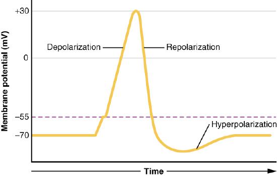
The signal generated by a motor neuron begins with opening of sodium channels in the membrane of the nerve cell body causing a rapid influx of sodium ions into the nerve cell. This step, called depolarization (Figure 3.42), triggers an electrochemical signal - the action potential. Remember that the Na+/K+ ATPase has created a large sodium gradient, so sodium ions rush into the cell when sodium channels open. After the initial depolarization, potassium channel gates, responding to the depolarization, open, allowing potassium ions to rapidly diffuse out of the cell (remember K+ ions are more abundant inside of the cell). This phase is called the repolarization phase and during it, the sodium gates close.
The rapid exit of potassium ions causes the voltage difference to “overshoot” the resting potential and potassium gates close. This followed by the so-called refractory period, when the Na+/K+ ATPase begins its work to re-establish the original conditions by pumping sodium ions out and potassium ions into the nerve cell. Eventually, the system recovers and the resting potential is re-established. The initiating end of the nerve cell is then ready for another signal.
Propagation of action potential
What we have described here is only the initiation of the nerve signal in one part of the nerve cell. For the signal to be received, the action potential must travel the entirety of the length of the nerve cell (the axon) and cause a chemical signal to be released into the synaptic cleft to get to its target. Propagating the nerve signal (action potential) in the original nerve cell is the function of all of the rest of the gated ion channels (Figure 3.43) positioned on the sides of the nerve cell. The sodium and potassium gates involved in propagation of the signal all act in response to voltage changes created by the electrochemical gradient moving down the nerve cell (Figure 3.44). Remember that opening of the initial gates at initiation of the signal created an influx of sodium ions and an efflux of potassium ions.
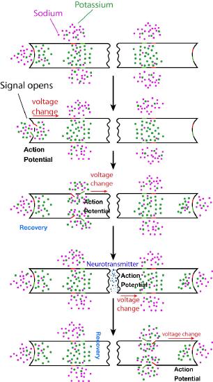
Moving signal
This chemical and electrical change that creates the action potential leaves the end of the nerve cell where it started and travels down the axon towards the other end of the nerve cell. Along the way, it encounters more sodium and potassium gated channels. In each case, these respond simply to the voltage change of the action potential and open and close, exactly in the same way the gates opened to start the signal. Thus, a rapid wave of increasing sodium ions and decreasing potassium ions moves along the nerve cell, propagated (and amplified) by gates opening and closing as the ions and charges move down the nerve cell. Eventually, the ionic tidal wave reaches the end of the nerve cell (axon terminal) facing the synaptic cleft.
Crossing the synaptic cleft
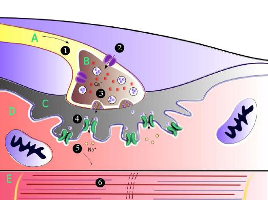
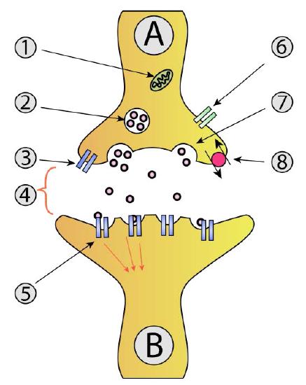
For the signal to be received by the intended target (postsynaptic cell) from the originating neuron (presynaptic neuron), it must cross the synaptic cleft and stimulate the neighboring cell (Figure 3.45). Communicating information across a synaptic cleft is the job of neurotransmitters. These are small molecules synthesized in nerve cells that are packaged in membrane vesicles called synaptic vesicles in the nerve cell. Neurotransmitters come in all shapes and chemical forms, from small chemicals like acetylcholine to peptides like neuropeptide Y. The most abundant neurotransmitter is glutamate, which acts at over 90% of the synapses in the human brain.
Movie 3.2 - Movement of an action potential down a nerve cell - Wikipedia
Into the cleft
As the action potential in the presynaptic neuron approaches the axon terminus, synaptic vesicles begin to fuse with the membrane and their neurotransmitter contents spill into the synaptic cleft. Once in the cleft, the neurotransmitters diffuse, some of them reaching receptors on the postsynaptic cell. Binding of the neurotransmitter to the receptors on the membrane of the postsynaptic cell stimulates a response.
For motor neurons, the postsynaptic cell will be a muscle cell, and the response will be muscle contraction/relaxation. At this point, the originating nerve cell has done its job and communicated its information to its immediate target. If the postsynaptic cell is a nerve cell, the process repeats in that cell until it gets to its destination.