
Structure Determination of Alkanes Using 13C-NMR and H-NMR
- Page ID
- 457128
This page is a draft and is under active development.
\( \newcommand{\vecs}[1]{\overset { \scriptstyle \rightharpoonup} {\mathbf{#1}} } \)
\( \newcommand{\vecd}[1]{\overset{-\!-\!\rightharpoonup}{\vphantom{a}\smash {#1}}} \)
\( \newcommand{\dsum}{\displaystyle\sum\limits} \)
\( \newcommand{\dint}{\displaystyle\int\limits} \)
\( \newcommand{\dlim}{\displaystyle\lim\limits} \)
\( \newcommand{\id}{\mathrm{id}}\) \( \newcommand{\Span}{\mathrm{span}}\)
( \newcommand{\kernel}{\mathrm{null}\,}\) \( \newcommand{\range}{\mathrm{range}\,}\)
\( \newcommand{\RealPart}{\mathrm{Re}}\) \( \newcommand{\ImaginaryPart}{\mathrm{Im}}\)
\( \newcommand{\Argument}{\mathrm{Arg}}\) \( \newcommand{\norm}[1]{\| #1 \|}\)
\( \newcommand{\inner}[2]{\langle #1, #2 \rangle}\)
\( \newcommand{\Span}{\mathrm{span}}\)
\( \newcommand{\id}{\mathrm{id}}\)
\( \newcommand{\Span}{\mathrm{span}}\)
\( \newcommand{\kernel}{\mathrm{null}\,}\)
\( \newcommand{\range}{\mathrm{range}\,}\)
\( \newcommand{\RealPart}{\mathrm{Re}}\)
\( \newcommand{\ImaginaryPart}{\mathrm{Im}}\)
\( \newcommand{\Argument}{\mathrm{Arg}}\)
\( \newcommand{\norm}[1]{\| #1 \|}\)
\( \newcommand{\inner}[2]{\langle #1, #2 \rangle}\)
\( \newcommand{\Span}{\mathrm{span}}\) \( \newcommand{\AA}{\unicode[.8,0]{x212B}}\)
\( \newcommand{\vectorA}[1]{\vec{#1}} % arrow\)
\( \newcommand{\vectorAt}[1]{\vec{\text{#1}}} % arrow\)
\( \newcommand{\vectorB}[1]{\overset { \scriptstyle \rightharpoonup} {\mathbf{#1}} } \)
\( \newcommand{\vectorC}[1]{\textbf{#1}} \)
\( \newcommand{\vectorD}[1]{\overrightarrow{#1}} \)
\( \newcommand{\vectorDt}[1]{\overrightarrow{\text{#1}}} \)
\( \newcommand{\vectE}[1]{\overset{-\!-\!\rightharpoonup}{\vphantom{a}\smash{\mathbf {#1}}}} \)
\( \newcommand{\vecs}[1]{\overset { \scriptstyle \rightharpoonup} {\mathbf{#1}} } \)
\(\newcommand{\longvect}{\overrightarrow}\)
\( \newcommand{\vecd}[1]{\overset{-\!-\!\rightharpoonup}{\vphantom{a}\smash {#1}}} \)
\(\newcommand{\avec}{\mathbf a}\) \(\newcommand{\bvec}{\mathbf b}\) \(\newcommand{\cvec}{\mathbf c}\) \(\newcommand{\dvec}{\mathbf d}\) \(\newcommand{\dtil}{\widetilde{\mathbf d}}\) \(\newcommand{\evec}{\mathbf e}\) \(\newcommand{\fvec}{\mathbf f}\) \(\newcommand{\nvec}{\mathbf n}\) \(\newcommand{\pvec}{\mathbf p}\) \(\newcommand{\qvec}{\mathbf q}\) \(\newcommand{\svec}{\mathbf s}\) \(\newcommand{\tvec}{\mathbf t}\) \(\newcommand{\uvec}{\mathbf u}\) \(\newcommand{\vvec}{\mathbf v}\) \(\newcommand{\wvec}{\mathbf w}\) \(\newcommand{\xvec}{\mathbf x}\) \(\newcommand{\yvec}{\mathbf y}\) \(\newcommand{\zvec}{\mathbf z}\) \(\newcommand{\rvec}{\mathbf r}\) \(\newcommand{\mvec}{\mathbf m}\) \(\newcommand{\zerovec}{\mathbf 0}\) \(\newcommand{\onevec}{\mathbf 1}\) \(\newcommand{\real}{\mathbb R}\) \(\newcommand{\twovec}[2]{\left[\begin{array}{r}#1 \\ #2 \end{array}\right]}\) \(\newcommand{\ctwovec}[2]{\left[\begin{array}{c}#1 \\ #2 \end{array}\right]}\) \(\newcommand{\threevec}[3]{\left[\begin{array}{r}#1 \\ #2 \\ #3 \end{array}\right]}\) \(\newcommand{\cthreevec}[3]{\left[\begin{array}{c}#1 \\ #2 \\ #3 \end{array}\right]}\) \(\newcommand{\fourvec}[4]{\left[\begin{array}{r}#1 \\ #2 \\ #3 \\ #4 \end{array}\right]}\) \(\newcommand{\cfourvec}[4]{\left[\begin{array}{c}#1 \\ #2 \\ #3 \\ #4 \end{array}\right]}\) \(\newcommand{\fivevec}[5]{\left[\begin{array}{r}#1 \\ #2 \\ #3 \\ #4 \\ #5 \\ \end{array}\right]}\) \(\newcommand{\cfivevec}[5]{\left[\begin{array}{c}#1 \\ #2 \\ #3 \\ #4 \\ #5 \\ \end{array}\right]}\) \(\newcommand{\mattwo}[4]{\left[\begin{array}{rr}#1 \amp #2 \\ #3 \amp #4 \\ \end{array}\right]}\) \(\newcommand{\laspan}[1]{\text{Span}\{#1\}}\) \(\newcommand{\bcal}{\cal B}\) \(\newcommand{\ccal}{\cal C}\) \(\newcommand{\scal}{\cal S}\) \(\newcommand{\wcal}{\cal W}\) \(\newcommand{\ecal}{\cal E}\) \(\newcommand{\coords}[2]{\left\{#1\right\}_{#2}}\) \(\newcommand{\gray}[1]{\color{gray}{#1}}\) \(\newcommand{\lgray}[1]{\color{lightgray}{#1}}\) \(\newcommand{\rank}{\operatorname{rank}}\) \(\newcommand{\row}{\text{Row}}\) \(\newcommand{\col}{\text{Col}}\) \(\renewcommand{\row}{\text{Row}}\) \(\newcommand{\nul}{\text{Nul}}\) \(\newcommand{\var}{\text{Var}}\) \(\newcommand{\corr}{\text{corr}}\) \(\newcommand{\len}[1]{\left|#1\right|}\) \(\newcommand{\bbar}{\overline{\bvec}}\) \(\newcommand{\bhat}{\widehat{\bvec}}\) \(\newcommand{\bperp}{\bvec^\perp}\) \(\newcommand{\xhat}{\widehat{\xvec}}\) \(\newcommand{\vhat}{\widehat{\vvec}}\) \(\newcommand{\uhat}{\widehat{\uvec}}\) \(\newcommand{\what}{\widehat{\wvec}}\) \(\newcommand{\Sighat}{\widehat{\Sigma}}\) \(\newcommand{\lt}{<}\) \(\newcommand{\gt}{>}\) \(\newcommand{\amp}{&}\) \(\definecolor{fillinmathshade}{gray}{0.9}\)Introduction.
In your text, the following table was included to illustrate the similarities in the physical properties of four alkanes, all with six carbon atoms. If you were provided with four unlabeled vials, each containing one of the compounds listed, could you unambiguously determine which is which?
| Table 1-1. Properties of Selected Alkanes Having Six Carbon Atoms | ||||
| Acyclic Alkanes | Cyclic Alkanes | |||
|
hexane |
2-methylpentane |
cyclohexane |
methylcyclopentane |
|
|
Molecular Formula |
C6H14 |
C6H14 |
C6H12 |
C6H12 |
| Line Diagram | ||||
|
Description |
colorless liquid |
colorless liquid |
colorless liquid |
colorless liquid |
|
Density (g/mL) |
0.655 |
0.653 |
0.778 |
0.749 |
|
Boiling point (°C) |
69 |
60 |
81 |
72 |
|
Solubility in water (ppm = mg/L) |
12 |
14 |
58 |
45 |
Given the information in the table, yes, it is theoretically possible to do so, but it would be fairly labor intensive and not as straightforward as you might think. For example, it would be difficult, for purely practical reasons, to distinguish between hexane and 2-methylhexane on the basis of their densities. Why? The masses of 100.0 mL samples of them would only differ by 0.2 grams, assuming there is absolutely no error in the volume measurement. But if the uncertainty of volume measurements were 0.1% (typical for volumetric flasks), then it is possible that the mass readings could be identical, or even that the 2-methylpentane sample would have a slightly greater mass than the hexane. You could try to determine which was which by experimentally measuring their boiling point, but that requires large enough samples to obtain reliable temperature measurements.
There is, instead, a much more elegant and unambiguous method to make such determinations. Nuclear magnetic resonance spectroscopy (NMR), is a powerful technique that is widely used to determine the structures of organic compounds. It does so by measuring the absorption of radio frequency electromagnetic radiation in the presence of a strong magnetic field and can distinguish atoms in different chemical environments. What does that mean? You can think of the chemical environment of an atom as being its surroundings in the molecule: specifically what other atoms it is connected to and how those are, in turn, connected to other atoms. Different elements require slightly different hardware to perform NMR and the techniques are named to reflect this; to examine carbon atoms, carbon-13 NMR (13C NMR) is used (the “13” indicates the specific isotope used; we’ll discuss isotopes in Chapter 3). We’ll start our discussion of NMR by focusing on 13C NMR then move on to hydrogen NMR (H NMR).

To illustrate how to use 13C-NMR spectra to determine molecular structure, we’ll take hexane as an example. The line diagram of hexane is shown above, and each carbon is labeled. Starting with the terminal methyl on the left, carbon 1, it “feels” the presence of carbon 2 in addition to the three hydrogen atoms it is bound to but which are not shown. Carbon 2, in turn, “feels” the presence of carbons 1 and 3, as well as its own two hydrogen atoms. This is a distinctly different set of interactions than those felt by carbon 1 and, as a result, these two carbons are in different chemical environments and they can be distinguished spectroscopically by 13C NMR.
What about Carbons 2 and 3? They are both connected to two carbon atoms and two hydrogen atoms - does this make them equivalent? No, it doesn’t. They have different chemical environments because carbon 2 is bound to a methyl group (carbon 1) and a methylene group (carbon 3; “methylene” is the term used for a -CH2- group), while carbon 3 is bound to two methylene groups (carbons 2 and 4). Thus carbons 2 and 3 are also distinguishable spectroscopically. Another way of thinking about these two atoms is that carbon 2 is bound to a methyl group and an n-butyl group (carbons 3-6), while carbon 3 is bound to an ethyl group (carbons 1 and 2) and an n-propyl group (carbons 4-6).
Moving on to carbons 3 and 4, these are equivalent and cannot be distinguished by NMR. Why? They are identical in that both are connected to an ethyl group and an n-propyl group. Prove that to yourself using the structure above. Likewise, carbons 2 and 5 are indistinguishable from each other, as are carbons 1 and 6. Hexane, therefore, will exhibit 3 signals in the 13C NMR spectrum (shown below), one for each set of indistinguishable carbon atoms, often called “unique” carbon atoms.
NMR spectra are presented as signal intensity as a function of magnetic field strength, expressed as ppm. The three peaks in the spectrum above are very sharp spikes at roughly 17, 22.5 and 32 ppm. At this point you need not worry about which signal corresponds with which carbon, although the theory that explains it is very well developed. Focus rather on the number of peaks and how that relates to the molecular structure.

Figure 1. 13C NMR spectrum of hexane (simulation obtained using software at nmrdb.org); the number of carbon atoms responsible for each peak are shown in red (more information about peak intensities is provided below).
From the example above, you may have a suspicion that you can predict if carbon atoms will be equivalent by symmetry. This is indeed the case, although sometimes it is tricky to recognize the symmetry. In the case of hexane, the left and right sides of the structure are equivalent by symmetry, specifically by a 180° rotation around the midpoint of the molecule as illustrated below. The image on the right is indistinguishable from the image on the left (other than the labels, which also rotated). A rotation or reflection of a structure that results in a new spatial orientation that is indistinguishable from the original is called a symmetry operation. The specific operation, that is, a rotation or a reflection, is called a symmetry element. This leads us to our first general principle when thinking about NMR spectra:
- atoms that exchange places as a result of a symmetry operation (or combination of symmetry operations) have identical chemical environments and are indistinguishable in NMR spectroscopy.

A very important caveat applies to using symmetry, however. Specifically, the symmetry operation be operative on the 3-dimensional molecule, not just a two-dimensional representation of it. In the example above, the operation works because the six carbon atoms of hexane line in the same plane in the actual structure, meaning that no significant distortion is introduced when drawing it in two dimensions. More often than not, this is not the case, so using a ball-and-stick model kit can be a big help in determining which, if any, symmetry elements a molecule possesses.
Using the above principle, let’s examine how many peaks we might expect for 2-methylpentane, shown below.


There is no rotational symmetry in this structure, but is there a mirror plane? It’s not obvious from the line structure above, but there is. Recall from your reading that line drawings are highly abstracted constructions that convey the essential structural elements of a molecule but necessarily involves some distortion. If you were to have a model kit and build the molecule, you could arrange the atoms in such a way that a mirror plane bisects the molecule, as illustrated in the ball-and-stick illustration at right. When viewed this way, it is apparent that carbons 1 and 6 are equivalent because they reflect into each other, while carbons 2, 3, 4 and 5, which lie in the mirror plane itself and don't reflect into other atoms, are all unique. We would expect, therefore that the NMR spectrum of 2-methyl pentane would have 5 total peaks. The spectrum below confirms this prediction.

Figure 2. 13C NMR spectrum of 2-methypentane (simulation obtained using software at nmrdb.org)
The spectrum above reveals another way NMR spectra can be used to help determine the structure of a compound: the peaks sizes. The intensity of the signals on NMR spectra, which are usually quantified by the integrated areas under the peaks, are directly proportional to the number of atoms that give rise to the signal. Thus in the above spectrum, there are one large peak and four smaller ones, with integrated areas in the ratio of 2:1:1:1:1, reflecting the number of atoms in the molecule that are responsible for each signal. In the simulations generated for this lab, the numbers in red at the top of the spectrum indicate the number of carbon atoms that are responsible for each peak. In practice, however, the are sometimes expressed as a percentage of the sum of all the peak areas. If that were they were expressed that way in the above example, they would be reported as 33.3:16.7:16.7:16.7:16.7 (corresponding to two-sixths and one-sixth the total integrated area). It would be up to you to translate these values into numbers of carbon atoms in the molecule.
Let’s now predict the 13C NMR spectra for the two cyclic alkanes from Table 1-1. Cyclohexane is a highly symmetric molecule, having both rotational and mirror symmetry. As illustrated below, if the molecule were completely flat (which it's not [1]), a 60° rotation would shift carbon 1 into the position of carbon 2, making them equivalent and at the same time making all of the other carbons equivalent to them as well (2 and 3 are equivalent, as are 3 and 4, etc). There should therefore only be 1 signal on the 13C NMR spectrum of cyclohexane. The spectrum below is in accord with our prediction (despite the fact that we distorted the structure bt "flattening it"; this shows that it is possible to make a correct prediction even if the logic employed is suspect).


Figure 3. 13C NMR spectrum of cyclohexane (simulation obtained using software at nmrdb.org).
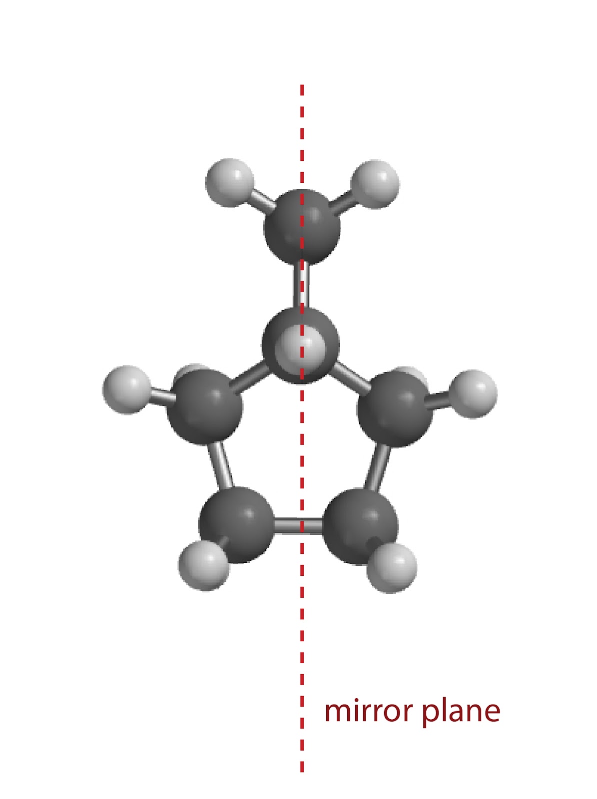


Figure 4. 13C NMR spectrum of methylcyclopentane (simulation obtained using software at nmrbd.org).
Group Exercise 1: Is the 13C NMR spectrum shown below obtained from 3-ethylpentane, or an isomer of this compound, 3-methylhexane? Explain your logic.

Group Exercise 2: For the compound that you didn’t choose above, predict the number of peaks in the 13C NMR spectrum. Simulate the spectrum by going to https://www.nmrdb.org/13c/index.shtml?v=v2.138.0; sketch the molecule in the workspace and then click “Predict Spectrum”. Do the number of peaks in the simulated spectrum match your prediction? If not, explain why.
Group Exercise 3: There are 5 isomers of hexane. Two of them give the same number of peaks on their respective 13C NMR spectra. What are these two structures? Can the peak sizes be used to distinguish these compounds using 13C NMR? Explain. Simulate the spectra to check your prediction.
Part 2: Hydrogen NMR
The third question above reveals one shortcoming to how we have been using 13C NMR thus far. Namely, there are many compounds that yield the same number of peaks on their 13C NMR spectra. What to do? [2] As the follow-up question above indicated, you can use the peak sizes to help determine the structure but this will not always work, especially if you don’t know anything about the structures.
Additional insight to molecular structure comes from performing NMR on the hydrogen atoms (1H NMR). H NMR spectra are often quite a bit more complicated than 13C NMR because the hydrogen atoms perturb the chemical environments of their nearest neighbors more than carbon atoms do. This leads to peak “splitting”; whereas in 13C NMR the carbon signals were all “singlets” meaning they consist of a single peak, peaks on the H NMR can be singlets, doublets, triplets, quartets, etc. Here’s an example: the H NMR spectrum of propane.

Figure 5: Simulated 1H NMR spectrum of propane (simulation obtained using software at nmrdb.org).
The 13C NMR of propane would have 2 peaks. (Why?) Similarly, the H NMR spectrum also has two signals, indicating that there are two sets of equivalent hydrogen atoms. But these signals don't appear as two single sharp peaks; the signal on the right has three peaks and is called a “triplet” and that on the left has seven peaks, called a “septet”. Their integrated areas have a 3:1 ratio (shown as 6 and 2 in the figure), meaning that the triplet is due to a set of six equivalent hydrogen atoms (shown in red, below) and the heptet is due to two equivalent hydrogen atoms (shown in blue).

As you can see, the six hydrogen atoms labeled in red, that is, those in the two methyl groups, give rise to the triplet. Does this mean methyl groups, because they have three hydrogen atoms each, are always triplets? Absolutely not! That is a common mistake that students make. There is a pattern to how hydrogen peaks are split, but it does not involve how many are on a given carbon. Instead, the logic of the splitting patterns can be summarized as follows:
-
Hydrogen atoms on equivalent carbon atoms are usually equivalent [3]; and
-
A signal from a given set of equivalent hydrogen atoms will be “split” according to the formula: number of peaks = n + 1, where n is the number of hydrogen atoms on adjacent carbon atoms, provided they are not part of that equivalent set (equivalent hydrogens can’t split their own signal).
The first point above is fairly straightforward, and the figure of propane illustrates it nicely. The two methyl carbon atoms are equivalent by symmetry (both rotation and reflection), thus the six hydrogen atoms attached to them are also equivalent with each other.
The second point is a bit more involved, Let’s examine the signal due to the 6 methyl hydrogen atoms. Looking at either methyl group, it is adjacent to a carbon atom that bears two hydrogen atoms. The value of n in this case is therefore 2, yielding a triplet according to the n + 1 formula. This is what we observe. As for the two hydrogen atoms on the central carbon, that carbon is adjacent to two carbon atoms bearing a total of six hydrogen atoms; thus n = 6 and the result is the observed septet. The last part of point 2 is not relevant to this example, but it is to the one below.
A few more examples may be helpful. Let’s consider the two isomers of butane, beginning with the unbranched form, shown below.

Carbon atoms 1 and 4 are equivalent by symmetry (how so?), making the six hydrogen atoms bonded to them equivalent as well. Similarly, carbon atoms 2 and 3 are also equivalent, making the four hydrogen atoms bound to them equivalent. At this point we predict two signals on the H NMR, having integrated areas in the ratio of 3:2. As for the splitting, each methyl group is adjacent to a methylene, having two hydrogen atoms. Therefore the larger peak should be split into a triplet (because n = 2). If you look at carbon 2, you can see that it is adjacent to two carbon atoms, 1 and 3, and they have a total of 5 hydrogen atoms. But the signal is not split into a sextet. Why? Because hydrogen atoms that are equivalent to each other don’t split each other’s signal. Carbons 2 and 3 are equivalent, so the hydrogen atoms on carbon 3 don’t count toward the value of n when considering how the signal of the hydrogens of that group are split; only the hydrogen atoms of carbon 1 split the signal of the hydrogen atoms on carbon 2. As such, the value of n for the hydrogen atoms on carbon 2 (and 3) is 3, making the signal appear as a quartet. To summarize, the H NMR of butane will have a triplet and a quartet with areas in the ratio of 3:2, respectively. The simulated spectrum shows this, albeit with some additional minor features that arise from a more sophisticated treatment. Nevertheless, the major features as predicted above are indeed observed.

Figure 6: Simulated 1H NMR spectrum of butane (simulation obtained using software at nmrdb.org).
 The structure of 2-methylpropane is shown at left. It looks like the hydrogen on carbon 2 disrupts the three-fold rotational symmetry of the molecule that would otherwise be present, but this an artifact of the 2-D representation. When the 3-D molecule is situated as in the ball and stick model at right, the blue hydrogen is situated directly in front of carbon 2 (think of it as coming out of the page) and the rotational symmetry of the molecule is much more evident. Thus carbon atoms 1, 3 and 4 are equivalent because they can rotate into each other. They have a total of nine hydrogen atoms (three of which are hidden behind carbon atoms in the ball and stick figure) that are also equivalent (shown in red at left). Carbon 2 is unique and has only one hydrogen atom. We therefore expect to see two signals that have integrated areas in the ratio of 9:1.

How will the signals be split? This is a straightforward example of the n + 1 rule. The peak due to the nine equivalent hydrogen atoms will be split into a doublet because they are all adjacent to carbon 2 which has only one hydrogen. The hydrogen on carbon 2, however, is adjacent to nine hydrogen atoms, meaning it will be split into ten peaks (called a "decuplet"). When the signal is split into such a large number of peaks, however, it is often referred to as a “multiplet”. The simulated spectrum is shown below, but it is not possible to see all ten peaks of the decuplet because some of them are too small on this scale.

Figure 6: Simulated 1H NMR spectrum of 2-methylpropane (simulation obtained using software at nmrdb.org).
Group Exercise 4: Shown below are the H NMR spectra of the two isomers of hexane in Exercise 3 that have the same number of peaks in their 13C NMR spectra. Determine which structures correspond to which spectra.
-
Note: this spectrum the following splitting patterns (and integrated areas): doublet (3 H), triplet (6 H), quintet (4 H), and an octet (1 H).

-
Note: this spectrum features a singlet (9 H), a triplet (3 H) and a quartet (2 H).

Group Exercise 5. The 13C NMR and H NMR spectra of a compound having the formula C7H16 are shown below. Determine the structure of the compound.
13C NMR:

H NMR: Note this spectrum features a singlet (6 H), a triplet (6 H), and a quartet (2 H).

Individual Assignment: You will be assigned an isomer of octane. Predict the 13C NMR and H NMR. Record your predictions in your notebook and explain your logic. Then simulate the spectra and comment on the accuracy of your predictions. Print the spectra and affix them in your notebook.
Footnotes.
[1] The molecule is not flat at all. In actuality carbon 1 and 6 do not rotate into each other via 60º rotations. The molecule is "puckered" in such a way that carbons 1, 3 and 5 are equivalent by way of 120º rotations, as are carbons 2, 4 and 6. It looks a bit like a crown, with alternating carbon atoms being above and below the avaerage plane of the molecule. as it turns out, carbons 1 and 6 (and all adjacent pairs of carbon atoms) are equivalent by a 60º degree rotation followed a reflection; this combination of symmetry elements is called an "improper axis of rotation." That said, for our current purposes, treating the molecular as planar allows us to correctly predict the spectrum without getting bogged down in details of symmetry.
[2]. We have not focused at all on the position of the peaks along the horizontal axis, called the “chemical shift”. The chemical shift reveals important structural information that can also be used to determine the structure of a compound. As a result, even if two compounds have 4 peaks, the spectra can still be used to determine which compound is which.
[3]. There are some exceptions to this “rule”, usually found in cyclic structures or others where rotation around bonds is highly constrained.