Basic Theory
- Page ID
- 272756
\( \newcommand{\vecs}[1]{\overset { \scriptstyle \rightharpoonup} {\mathbf{#1}} } \)
\( \newcommand{\vecd}[1]{\overset{-\!-\!\rightharpoonup}{\vphantom{a}\smash {#1}}} \)
\( \newcommand{\id}{\mathrm{id}}\) \( \newcommand{\Span}{\mathrm{span}}\)
( \newcommand{\kernel}{\mathrm{null}\,}\) \( \newcommand{\range}{\mathrm{range}\,}\)
\( \newcommand{\RealPart}{\mathrm{Re}}\) \( \newcommand{\ImaginaryPart}{\mathrm{Im}}\)
\( \newcommand{\Argument}{\mathrm{Arg}}\) \( \newcommand{\norm}[1]{\| #1 \|}\)
\( \newcommand{\inner}[2]{\langle #1, #2 \rangle}\)
\( \newcommand{\Span}{\mathrm{span}}\)
\( \newcommand{\id}{\mathrm{id}}\)
\( \newcommand{\Span}{\mathrm{span}}\)
\( \newcommand{\kernel}{\mathrm{null}\,}\)
\( \newcommand{\range}{\mathrm{range}\,}\)
\( \newcommand{\RealPart}{\mathrm{Re}}\)
\( \newcommand{\ImaginaryPart}{\mathrm{Im}}\)
\( \newcommand{\Argument}{\mathrm{Arg}}\)
\( \newcommand{\norm}[1]{\| #1 \|}\)
\( \newcommand{\inner}[2]{\langle #1, #2 \rangle}\)
\( \newcommand{\Span}{\mathrm{span}}\) \( \newcommand{\AA}{\unicode[.8,0]{x212B}}\)
\( \newcommand{\vectorA}[1]{\vec{#1}} % arrow\)
\( \newcommand{\vectorAt}[1]{\vec{\text{#1}}} % arrow\)
\( \newcommand{\vectorB}[1]{\overset { \scriptstyle \rightharpoonup} {\mathbf{#1}} } \)
\( \newcommand{\vectorC}[1]{\textbf{#1}} \)
\( \newcommand{\vectorD}[1]{\overrightarrow{#1}} \)
\( \newcommand{\vectorDt}[1]{\overrightarrow{\text{#1}}} \)
\( \newcommand{\vectE}[1]{\overset{-\!-\!\rightharpoonup}{\vphantom{a}\smash{\mathbf {#1}}}} \)
\( \newcommand{\vecs}[1]{\overset { \scriptstyle \rightharpoonup} {\mathbf{#1}} } \)
\( \newcommand{\vecd}[1]{\overset{-\!-\!\rightharpoonup}{\vphantom{a}\smash {#1}}} \)
\(\newcommand{\avec}{\mathbf a}\) \(\newcommand{\bvec}{\mathbf b}\) \(\newcommand{\cvec}{\mathbf c}\) \(\newcommand{\dvec}{\mathbf d}\) \(\newcommand{\dtil}{\widetilde{\mathbf d}}\) \(\newcommand{\evec}{\mathbf e}\) \(\newcommand{\fvec}{\mathbf f}\) \(\newcommand{\nvec}{\mathbf n}\) \(\newcommand{\pvec}{\mathbf p}\) \(\newcommand{\qvec}{\mathbf q}\) \(\newcommand{\svec}{\mathbf s}\) \(\newcommand{\tvec}{\mathbf t}\) \(\newcommand{\uvec}{\mathbf u}\) \(\newcommand{\vvec}{\mathbf v}\) \(\newcommand{\wvec}{\mathbf w}\) \(\newcommand{\xvec}{\mathbf x}\) \(\newcommand{\yvec}{\mathbf y}\) \(\newcommand{\zvec}{\mathbf z}\) \(\newcommand{\rvec}{\mathbf r}\) \(\newcommand{\mvec}{\mathbf m}\) \(\newcommand{\zerovec}{\mathbf 0}\) \(\newcommand{\onevec}{\mathbf 1}\) \(\newcommand{\real}{\mathbb R}\) \(\newcommand{\twovec}[2]{\left[\begin{array}{r}#1 \\ #2 \end{array}\right]}\) \(\newcommand{\ctwovec}[2]{\left[\begin{array}{c}#1 \\ #2 \end{array}\right]}\) \(\newcommand{\threevec}[3]{\left[\begin{array}{r}#1 \\ #2 \\ #3 \end{array}\right]}\) \(\newcommand{\cthreevec}[3]{\left[\begin{array}{c}#1 \\ #2 \\ #3 \end{array}\right]}\) \(\newcommand{\fourvec}[4]{\left[\begin{array}{r}#1 \\ #2 \\ #3 \\ #4 \end{array}\right]}\) \(\newcommand{\cfourvec}[4]{\left[\begin{array}{c}#1 \\ #2 \\ #3 \\ #4 \end{array}\right]}\) \(\newcommand{\fivevec}[5]{\left[\begin{array}{r}#1 \\ #2 \\ #3 \\ #4 \\ #5 \\ \end{array}\right]}\) \(\newcommand{\cfivevec}[5]{\left[\begin{array}{c}#1 \\ #2 \\ #3 \\ #4 \\ #5 \\ \end{array}\right]}\) \(\newcommand{\mattwo}[4]{\left[\begin{array}{rr}#1 \amp #2 \\ #3 \amp #4 \\ \end{array}\right]}\) \(\newcommand{\laspan}[1]{\text{Span}\{#1\}}\) \(\newcommand{\bcal}{\cal B}\) \(\newcommand{\ccal}{\cal C}\) \(\newcommand{\scal}{\cal S}\) \(\newcommand{\wcal}{\cal W}\) \(\newcommand{\ecal}{\cal E}\) \(\newcommand{\coords}[2]{\left\{#1\right\}_{#2}}\) \(\newcommand{\gray}[1]{\color{gray}{#1}}\) \(\newcommand{\lgray}[1]{\color{lightgray}{#1}}\) \(\newcommand{\rank}{\operatorname{rank}}\) \(\newcommand{\row}{\text{Row}}\) \(\newcommand{\col}{\text{Col}}\) \(\renewcommand{\row}{\text{Row}}\) \(\newcommand{\nul}{\text{Nul}}\) \(\newcommand{\var}{\text{Var}}\) \(\newcommand{\corr}{\text{corr}}\) \(\newcommand{\len}[1]{\left|#1\right|}\) \(\newcommand{\bbar}{\overline{\bvec}}\) \(\newcommand{\bhat}{\widehat{\bvec}}\) \(\newcommand{\bperp}{\bvec^\perp}\) \(\newcommand{\xhat}{\widehat{\xvec}}\) \(\newcommand{\vhat}{\widehat{\vvec}}\) \(\newcommand{\uhat}{\widehat{\uvec}}\) \(\newcommand{\what}{\widehat{\wvec}}\) \(\newcommand{\Sighat}{\widehat{\Sigma}}\) \(\newcommand{\lt}{<}\) \(\newcommand{\gt}{>}\) \(\newcommand{\amp}{&}\) \(\definecolor{fillinmathshade}{gray}{0.9}\)Description
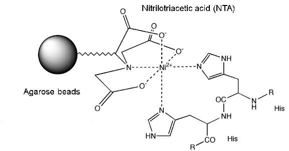
According to the International Union of Pure and Applied Chemistry, affinity chromatography is defined as a liquid chromatographic technique that makes use of a "biological interaction" for the separation and analysis of specific analytes within a sample. For example, a protein that binds metals such as nickel can be purified in the presence of other non-specific proteins using a resin containing immobilized nickel. A protein can bind nickel because of the presence of amino acids such as histidine positioned in a specific manner, which contains imidazole functionality that can co-ordinate nickel ion as shown in the figure below.

The biomolecule of interest interacts reversibly with a specific ligand bound to a matrix allowing for a specific binding on the matrix in the presence of other contaminants and later elution of the bound biomolecule. Using this method the biomolecule can be purified in a single step, with efficient recovery and high purity.
There are certain specific requirements for an affinity chromatography that must be met. These requirements are as follows:
- A biospecific ligand that can be covalently attached to a chromatography matrix.
- The bound ligand must be able to bind the target biomolecule specifically.
- The binding between the ligand and target molecule must be reversible to allow the target molecules to be removed in an active form.
What are the types of biological interactions used in affinity chromatography?
Some of the examples of types of interactions utilized in the affinity chromatographic purification include,
- Antigen : antibody
- Enzyme : substrate analogue
- Binding protein: Ligand
- Receptor : ligand
- Lectin : polysaccharide, glycoprotein
- Nucleic acid : complementary base sequence
- Hormone, vitamin : receptor, carrier protein.
- Glutathione : glutathione-S-transferase or GST fusion proteins.
- Metal ions : Poly (His) fusion proteins, native proteins with histidine or cysteine on their surfaces.
These biological interactions between ligand and target biomolecule can be a result of electrostatic or hydrophobic interactions, van der Waals' forces and/or hydrogen bonding. The elution of the target molecule from the matrix can be achieved either biospecifically using a competitive ligand, or non-specifically, by denaturing the biomoleule by changing the pH, ionic strength or polarity.
What are the Steps involved in Affinity Chromatography?
A movie demonstrating steps involved in affinity chromatography can be found in the link below developed by Amersham Biosciences,
Any component can be used as a ligand to purify its respective binding partner. The steps involved in a typical affinity chromatographic separation are as follows,
- The ligand is first covalently coupled to a matrix, such as agarose beads.
- The matrix is poured into a column.
- An impure mixture containing biomoleule of interest is loaded on the affinity column
- Biomolecules sieve through matrix of affinity beads and interact with affinity ligand. Molecules that do not bind to ligand elute from the column
- Wash off contaminant molecules that bind to ligand loosely
- Elute proteins that bind tightly to ligand and collect purified protein of interest using either a biospeciifc or nonspecific elution methods
- Biospecific – An inhibitor is added to the mobile phase (free ligand). Free ligand will compete for the solute
- Nonspecific – A reagent is added that denatures the solute. Once denatured, the solute is released from the ligand.
A diagram showing steps involved in affinity chromatographic separation and a typical elution profile is shown below.

How to use affinity purification when a specific ligand for your target biomolecule is not available?
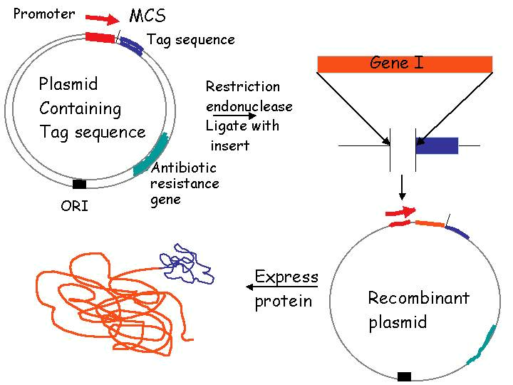
In general there are three groups of properties of the target molecule that are used in affinity chromatography. A simplest way is to take advantage of specific binding property of the biomolecule that you trying to purify, for example, enzyme active sites, receptor binding sites, antibody binding sites etc. These are used together with the natural ligand or an analogue of it. The other method is to use naturally occurring prosthetic groups like polysaccharides present on the proteins. You can bind ligand such as concanavalin A on the matric and used the interaction to perform affinity separation. However, this allows for only the separation of a group of biomolecules and may not be very efficient. The third way to purify the molecule is using affinity tags that are not present naturally on the biomolecule of your interest. For example, affinity tags such as Glutathione-S-Transferase (GST), six-histidine tag can be fused to molecules of your interest using genetic coupling techniques (Figure shown below). These tags then allow for your biomoleule of interest to bind to ligand followed by elution using appropriate conditions. This way of purification is most commonly used for purification of recombinant fusion proteins and it can be applied to most any biomolecule of interest.

How do you analyze purified biomolecules?
Depending upon the type and property of biomolecule that you are purifying various techniques can be applied to evaluate the success of your affinity method. There are two important factors that need to be evaluated after performing the affinity separation. One of them is the purity of the eluted biomoleucle and the other is its activity. Performing activity check is essential to ensure that purification procedure is mild on the biomoleule of interest and the biomolecule maintains its full activity. The activity determination is a property of the molecule you are working with it and can be evaluated using appropriate assay methods available.
Purity can be evaluated using gel electrophoresis techniques. For proteins sodium dodecyl sulfate polyacrylamide gel electrophorsis (SDS-PAGE) is employed. In SDS-PAGE, the solution of proteins to be analyzed is first mixed with SDS, an anionic detergent which denatures secondary and non–disulfide–linked tertiary structures of proteins. It also applies a negative charge to each protein in proportion to its mass. SDS linearizes all the proteins in a sample such that they can be separated on the basis of their molecular weight. For the SDS-PAGE analysis proteins are briefly heated to near boiling in the presence of a reducing agent, such as dithiothreitol (DTT) or 2-mercaptoethanol (beta-Mercaptoethanol/BME), which further denatures the proteins by reducing disulfide linkages. The molecular weight of the protein of interest is verified by running a protein molecular weight marker. A picture of a typical SDS-PAGE stained using Coomassie Blue can be seen below. Lane 1 show a molecular weight marker, Lane 1 and 2 show unpurified sample, and lane 3-6 show purified protein of interest.

More information on gel electrophoresis can be found at the following resources:


