
3.10: Carbon-13 Nuclear Magnetic Resonance
- Page ID
- 201105
\( \newcommand{\vecs}[1]{\overset { \scriptstyle \rightharpoonup} {\mathbf{#1}} } \)
\( \newcommand{\vecd}[1]{\overset{-\!-\!\rightharpoonup}{\vphantom{a}\smash {#1}}} \)
\( \newcommand{\id}{\mathrm{id}}\) \( \newcommand{\Span}{\mathrm{span}}\)
( \newcommand{\kernel}{\mathrm{null}\,}\) \( \newcommand{\range}{\mathrm{range}\,}\)
\( \newcommand{\RealPart}{\mathrm{Re}}\) \( \newcommand{\ImaginaryPart}{\mathrm{Im}}\)
\( \newcommand{\Argument}{\mathrm{Arg}}\) \( \newcommand{\norm}[1]{\| #1 \|}\)
\( \newcommand{\inner}[2]{\langle #1, #2 \rangle}\)
\( \newcommand{\Span}{\mathrm{span}}\)
\( \newcommand{\id}{\mathrm{id}}\)
\( \newcommand{\Span}{\mathrm{span}}\)
\( \newcommand{\kernel}{\mathrm{null}\,}\)
\( \newcommand{\range}{\mathrm{range}\,}\)
\( \newcommand{\RealPart}{\mathrm{Re}}\)
\( \newcommand{\ImaginaryPart}{\mathrm{Im}}\)
\( \newcommand{\Argument}{\mathrm{Arg}}\)
\( \newcommand{\norm}[1]{\| #1 \|}\)
\( \newcommand{\inner}[2]{\langle #1, #2 \rangle}\)
\( \newcommand{\Span}{\mathrm{span}}\) \( \newcommand{\AA}{\unicode[.8,0]{x212B}}\)
\( \newcommand{\vectorA}[1]{\vec{#1}} % arrow\)
\( \newcommand{\vectorAt}[1]{\vec{\text{#1}}} % arrow\)
\( \newcommand{\vectorB}[1]{\overset { \scriptstyle \rightharpoonup} {\mathbf{#1}} } \)
\( \newcommand{\vectorC}[1]{\textbf{#1}} \)
\( \newcommand{\vectorD}[1]{\overrightarrow{#1}} \)
\( \newcommand{\vectorDt}[1]{\overrightarrow{\text{#1}}} \)
\( \newcommand{\vectE}[1]{\overset{-\!-\!\rightharpoonup}{\vphantom{a}\smash{\mathbf {#1}}}} \)
\( \newcommand{\vecs}[1]{\overset { \scriptstyle \rightharpoonup} {\mathbf{#1}} } \)
\( \newcommand{\vecd}[1]{\overset{-\!-\!\rightharpoonup}{\vphantom{a}\smash {#1}}} \)
\(\newcommand{\avec}{\mathbf a}\) \(\newcommand{\bvec}{\mathbf b}\) \(\newcommand{\cvec}{\mathbf c}\) \(\newcommand{\dvec}{\mathbf d}\) \(\newcommand{\dtil}{\widetilde{\mathbf d}}\) \(\newcommand{\evec}{\mathbf e}\) \(\newcommand{\fvec}{\mathbf f}\) \(\newcommand{\nvec}{\mathbf n}\) \(\newcommand{\pvec}{\mathbf p}\) \(\newcommand{\qvec}{\mathbf q}\) \(\newcommand{\svec}{\mathbf s}\) \(\newcommand{\tvec}{\mathbf t}\) \(\newcommand{\uvec}{\mathbf u}\) \(\newcommand{\vvec}{\mathbf v}\) \(\newcommand{\wvec}{\mathbf w}\) \(\newcommand{\xvec}{\mathbf x}\) \(\newcommand{\yvec}{\mathbf y}\) \(\newcommand{\zvec}{\mathbf z}\) \(\newcommand{\rvec}{\mathbf r}\) \(\newcommand{\mvec}{\mathbf m}\) \(\newcommand{\zerovec}{\mathbf 0}\) \(\newcommand{\onevec}{\mathbf 1}\) \(\newcommand{\real}{\mathbb R}\) \(\newcommand{\twovec}[2]{\left[\begin{array}{r}#1 \\ #2 \end{array}\right]}\) \(\newcommand{\ctwovec}[2]{\left[\begin{array}{c}#1 \\ #2 \end{array}\right]}\) \(\newcommand{\threevec}[3]{\left[\begin{array}{r}#1 \\ #2 \\ #3 \end{array}\right]}\) \(\newcommand{\cthreevec}[3]{\left[\begin{array}{c}#1 \\ #2 \\ #3 \end{array}\right]}\) \(\newcommand{\fourvec}[4]{\left[\begin{array}{r}#1 \\ #2 \\ #3 \\ #4 \end{array}\right]}\) \(\newcommand{\cfourvec}[4]{\left[\begin{array}{c}#1 \\ #2 \\ #3 \\ #4 \end{array}\right]}\) \(\newcommand{\fivevec}[5]{\left[\begin{array}{r}#1 \\ #2 \\ #3 \\ #4 \\ #5 \\ \end{array}\right]}\) \(\newcommand{\cfivevec}[5]{\left[\begin{array}{c}#1 \\ #2 \\ #3 \\ #4 \\ #5 \\ \end{array}\right]}\) \(\newcommand{\mattwo}[4]{\left[\begin{array}{rr}#1 \amp #2 \\ #3 \amp #4 \\ \end{array}\right]}\) \(\newcommand{\laspan}[1]{\text{Span}\{#1\}}\) \(\newcommand{\bcal}{\cal B}\) \(\newcommand{\ccal}{\cal C}\) \(\newcommand{\scal}{\cal S}\) \(\newcommand{\wcal}{\cal W}\) \(\newcommand{\ecal}{\cal E}\) \(\newcommand{\coords}[2]{\left\{#1\right\}_{#2}}\) \(\newcommand{\gray}[1]{\color{gray}{#1}}\) \(\newcommand{\lgray}[1]{\color{lightgray}{#1}}\) \(\newcommand{\rank}{\operatorname{rank}}\) \(\newcommand{\row}{\text{Row}}\) \(\newcommand{\col}{\text{Col}}\) \(\renewcommand{\row}{\text{Row}}\) \(\newcommand{\nul}{\text{Nul}}\) \(\newcommand{\var}{\text{Var}}\) \(\newcommand{\corr}{\text{corr}}\) \(\newcommand{\len}[1]{\left|#1\right|}\) \(\newcommand{\bbar}{\overline{\bvec}}\) \(\newcommand{\bhat}{\widehat{\bvec}}\) \(\newcommand{\bperp}{\bvec^\perp}\) \(\newcommand{\xhat}{\widehat{\xvec}}\) \(\newcommand{\vhat}{\widehat{\vvec}}\) \(\newcommand{\uhat}{\widehat{\uvec}}\) \(\newcommand{\what}{\widehat{\wvec}}\) \(\newcommand{\Sighat}{\widehat{\Sigma}}\) \(\newcommand{\lt}{<}\) \(\newcommand{\gt}{>}\) \(\newcommand{\amp}{&}\) \(\definecolor{fillinmathshade}{gray}{0.9}\)After completing this section, you should be able to
- Determine the number of distinct C atoms in a molecule.
- Use the chemical shifts table to determine functional groups present in a molecule.
- Assign a chemical shift to each carbon in a given molecule.
The Basics of 13C-NMR spectroscopy
Unlike 1H-NMR signals, the area under a 13C-NMR signal cannot be used to determine the number of carbons to which it corresponds. This is because the signals for some types of carbons are inherently weaker than for other types – peaks corresponding to carbonyl carbons, for example, are much smaller than those for methyl or methylene (CH2) peaks. Peak integration is generally not useful in 13C-NMR spectroscopy, except when investigating molecules that have been enriched with 13C isotope.
The resonance frequencies of 13C nuclei are lower than those of protons in the same applied field - in a 7.05 Tesla instrument, protons resonate at about 300 MHz, while carbons resonate at about 75 MHz. This is fortunate, as it allows us to look at 13C signals using a completely separate 'window' of radio frequencies. This means you will only see the 13C nuclei in a 13C NMR experiment like in the 1H NMR experiments we just looked at, we only saw hydrogens. Just like in 1H-NMR, the standard used in 13C-NMR experiments to define the 0 ppm point is tetramethylsilane (TMS), although of course in 13C-NMR it is the signal from the four equivalent carbons in TMS that serves as the standard. Chemical shifts for 13C nuclei in organic molecules are spread out over a much wider range than for protons – up to 200 ppm for 13C compared to 12 ppm for protons (see Table 3 for a list of typical 13C-NMR chemical shifts). This is also fortunate, because it means that the signal from each carbon in a compound can almost always be seen as a distinct peak, without the overlapping that often plagues 1H-NMR spectra. The chemical shift of a 13C nucleus is influenced by essentially the same factors that influence a proton's chemical shift: bonds to electronegative atoms and diamagnetic anisotropy effects tend to shift signals downfield (higher resonance frequency). In addition, sp2 hybridization results in a large downfield shift. The 13C-NMR signals for carbonyl carbons are generally the furthest downfield (170-220 ppm), due to both sp2 hybridization and to the double bond to oxygen.
- How many sets of non-equivalent carbons are there in each of the molecules shown in exercise 5.1?
- How many sets of non-equivalent carbons are there in:
- toluene
- 2-pentanone
- para-xylene
- triclosan
(all structures are shown earlier in this chapter)
Solution
a
a) 8 signals (each carbon is different)
b) 11 signals (the two enantiotopic CH2CH3 groups are NMR-equivalent)
c) 6 signals (each carbon is different)
d) 16 signals (the fluorobenzene group only contributes 4 signals due to symmetry)
b
a) 5 signals
b) 5 signals
c) 3 signals

d) 6 signals
Because of the low natural abundance of 13C nuclei, it is very unlikely to find two 13C atoms near each other in the same molecule, and thus we do not see spin-spin coupling between neighboring carbons in a 13C-NMR spectrum. There is, however, heteronuclear coupling between 13C carbons and the hydrogens to which they are bound. Carbon-proton coupling constants are very large, on the order of 100 – 250 Hz. For clarity, chemists generally use a technique called broadband decoupling, which essentially 'turns off' C-H coupling, resulting in a spectrum in which all carbon signals are singlets. Below is the proton-decoupled13C-NMR spectrum of ethyl acetate, showing the expected four signals, one for each of the carbons.

One of the greatest advantages of 13C-NMR compared to 1H-NMR is the breadth of the spectrum - recall that carbons resonate from 0-220 ppm relative to the TMS standard, as opposed to only 0-12 ppm for protons. Because of this, 13C signals rarely overlap, and we can almost always distinguish separate peaks for each carbon, even in a relatively large compound containing carbons in very similar environments. In the proton spectrum of 1-heptanol, for example, only the signals for the alcohol proton (Ha) and the two protons on the adjacent carbon (Hb) are easily analyzed. The other proton signals overlap, making analysis difficult.

In the 13C spectrum of the same molecule, however, we can easily distinguish each carbon signal, and we know from this data that our sample has seven non-equivalent carbons. (Notice also that, as we would expect, the chemical shifts of the carbons get progressively smaller as they get farther away from the deshielding oxygen.)

This property of 13C-NMR makes it very helpful in the elucidation of larger, more complex structures.
13C NMR Chemical Shifts
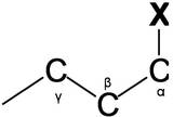
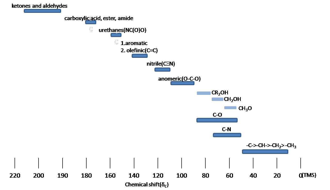
The Carbon NMR is used for determining functional groups using characteristic shift values. 13C chemical shifts are greatly affected by electronegative effects. If a H atom in an alkane is replaced by substituent X, electronegative atoms (O, N, halogen), 13C signals for nearby carbons shift downfield (left; increase in ppm) with the effect diminishing with distance from the electron withdrawing group. Figure 13.11.1 shows typical 13C chemical shift regions of the major chemical class.
Spin-Spin Splitting
Comparing the 1H NMR, there is a big difference thing in the 13C NMR. The 13C-13Cspin-spin splitting rarely exit between adjacent carbons because 13C is naturally lower abundant (1.1%)
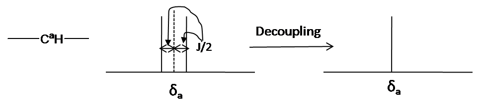
- 13C-1H Spin coupling: 13C-1H Spin coupling provides useful information about the number of protons attached a carbon atom. In case of one bond coupling (1JCH), -CH, -CH2, and CH3 have respectively doublet, triplet, quartets for the 13C resonances in the spectrum. However, 13C-1H Spin coupling has an disadvantage for 13C spectrum interpretation. 13C-1H Spin coupling is hard to analyze and reveal structure due to a forest of overlapping peaks that result from 100% abundance of 1H.
- Decoupling: Decoupling is the process of removing 13C-1H coupling interaction to simplify a spectrum and identify which pair of nuclei is involved in the J coupling. The decoupling 13C spectra shows only one peak(singlet) for each unique carbon in the molecule (Fig 13.11.2). Decoupling is performed by irradiating at the frequency of one proton with continuous low-power RF.
Summary
¹³C NMR (Carbon-13 Nuclear Magnetic Resonance) Spectroscopy is a powerful analytical technique used to study the structure and connectivity of organic molecules. Unlike proton NMR, which detects hydrogen nuclei, ¹³C NMR specifically targets the carbon nuclei within a molecule. In ¹³C NMR spectroscopy, carbon atoms resonate at characteristic frequencies based on their local chemical environment, which is influenced by neighboring atoms and functional groups. These resonances appear as peaks in the NMR spectrum, providing valuable information about the types of carbon atoms present and their surroundings. By analyzing the chemical shifts, intensities, and coupling patterns of these peaks, chemists can deduce crucial structural details such as the number and types of carbon atoms, their connectivity, and the presence of certain functional groups. This information is essential for determining the molecular structure and understanding chemical properties and reactivities.
After completing this section, you should be able to use data from 13C NMR spectra to distinguish between two (or more) possible structures for an unknown organic compound.
Features of a 13C NMR spectrum
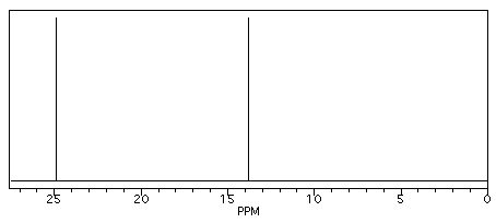
Butane shows two different peaks in the 13C NMR spectrum, below. Note that: the chemical shifts of these peaks are not very different from methane. The carbons in butane are in a similar environment to the one in methane.
- there are two distinct carbons in butane: the methyl, or CH3, carbon, and the methylene, or CH2, carbon.
- the methyl carbon absorbs slightly upfield, or at lower shift, around 10 ppm.
- the methylene carbon absorbs at slightly downfield, or at higher shift, around 20 ppm.
- other factors being equal, methylene carbons show up at slightly higher shift than methyl carbons.
In the 13C NMR spectrum of pentane (below), you can see three different peaks, even though pentane just contains methyl carbons and methylene carbons like butane. As far as the NMR spectrometer is concerned, pentane contains three different kinds of carbon, in three different environments. That result comes from symmetry.

Symmetry is an important factor in spectroscopy. Nature says:
- atoms that are symmetry-inequivalent can absorb at different shifts.
- atoms that are symmetry-equivalent must absorb at the same shift.
To learn about symmetry, take a model of pentane and do the following:
- make sure the model is twisted into the most symmetric shape possible: a nice "W".
- choose one of the methyl carbons to focus on.
- rotate the model 180 degrees so that you are looking at the same "W" but from the other side.
- note that the methyl you were focusing on has simply switched places with the other methyl group. These two carbons are symmetry-equivalent via two-fold rotation.
By the same process, you can see that the second and fourth carbons along the chain are also symmetry-equivalent. However, the middle carbon is not; it never switches places with the other carbons if you rotate the model. There are three different sets of inequivalent carbons; these three groups are not the same as each other according to symmetry.
Determine how many inequivalent carbons there are in each of the following compounds. How many peaks do you expect in each 13C NMR spectrum?

Practically speaking, there is only so much room in the spectrum from one end to the other. At some point, peaks can get so crowded together that you can't distinguish one from another. You might expect to see ten different peaks in eicosane, a twenty-carbon alkane chain, but when you look at the spectrum you can only see seven different peaks. That may be frustrating, because the experiment does not seem to agree with your expectation. However, you will be using a number of methods together to minimize the problem of misleading data.
Solution
a) Three inequivalent carbons/three peaks. There is a plane of symmetry that bisects the cyclohexene horizontally. The three different carbons are one of the alkene (C1), the CH2 next to alkene (C3) and C4.
b) Six inequivalent carbons/six peaks. The two methyl groups attached to the alkene are identical.
c) Four inequivalent carbons/four peaks. This molecule has a plane of symmetry through the molecule, including the methyl group. The two carbons adjacent to the methyl group are equivalent (C2 and C5). C3 and C4 are also equivalent.
d) Five inequivalent carbons/five peaks. This molecule has a plane of symmetry that passes through the ring carbon between the two methyl groups. The two methyl carbons are identical. The two ring carbons with the methyl groups attached are identical (C1 and C3). C4 and C6 are also equivalent.
e) Six inequivalent carbons/six peaks. The three methyl groups at the end of the molecule are equivalent.
f) Ten inequivalent carbons/ten peaks. There is no symmetry for the carbons in this molecule.
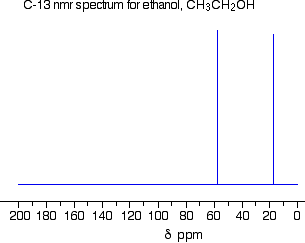
The 13C NMR spectrum for ethanol
This is a simple example of a 13C NMR spectrum. Don't worry about the scale for now - we'll look at that in a minute.
Note: The NMR spectra on this page have been produced from graphs taken from the Spectral Data Base System for Organic Compounds (SDBS) at the National Institute of Materials and Chemical Research in Japan.
There are two peaks because there are two different environments for the carbons. The carbon in the CH3 group is attached to 3 hydrogens and a carbon. The carbon in the CH2 group is attached to 2 hydrogens, a carbon and an oxygen. The two lines are in different places in the NMR spectrum because they need different external magnetic fields to bring them in to resonance at a particular radio frequency.
The 13C NMR spectrum for a more complicated compound
This is the 13C NMR spectrum for 1-methylethyl propanoate (also known as isopropyl propanoate or isopropyl propionate).
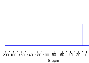
This time there are 5 lines in the spectrum. That means that there must be 5 different environments for the carbon atoms in the compound. Is that reasonable from the structure?

If you count the carbon atoms, there are 6 of them. So why only 5 lines? In this case, two of the carbons are in exactly the same environment. They are attached to exactly the same things. Look at the two CH3 groups on the right-hand side of the molecule.
You might reasonably ask why the carbon in the CH3 on the left is not also in the same environment. Just like the ones on the right, the carbon is attached to 3 hydrogens and another carbon. But the similarity is not exact - you have to chase the similarity along the rest of the molecule as well to be sure.
The carbon in the left-hand CH3 group is attached to a carbon atom which in turn is attached to a carbon with two oxygens on it - and so on down the molecule. That's not exactly the same environment as the carbons in the right-hand CH3 groups. They are attached to a carbon which is attached to a single oxygen - and so on down the molecule. We'll look at this spectrum again in detail on the next page - and look at some more similar examples as well. This all gets easier the more examples you look at.
For now, all you need to realize is that each line in a 13C NMR spectrum recognizes a carbon atom in one particular environment in the compound. If two (or more) carbon atoms in a compound have exactly the same environment, they will be represented by a single line.
You might wonder why all this works, since only about 1% of carbon atoms are 13C. These are the only ones picked up by this form of NMR. If you had a single molecule of ethanol, then the chances are only about 1 in 50 of there being one 13C atom in it, and only about 1 in 10,000 of both being 13C.
But you have got to remember that you will be working with a sample containing huge numbers of molecules. The instrument can pick up the magnetic effect of the 13C nuclei in the carbon of the CH3 group and the carbon of the CH2 group even if they are in separate molecules. There's no need for them to be in the same one.