
5.2.1: 5.2.1 Optional- other interesting lipids
- Page ID
- 347417

\( \newcommand{\vecs}[1]{\overset { \scriptstyle \rightharpoonup} {\mathbf{#1}} } \)
\( \newcommand{\vecd}[1]{\overset{-\!-\!\rightharpoonup}{\vphantom{a}\smash {#1}}} \)
\( \newcommand{\id}{\mathrm{id}}\) \( \newcommand{\Span}{\mathrm{span}}\)
( \newcommand{\kernel}{\mathrm{null}\,}\) \( \newcommand{\range}{\mathrm{range}\,}\)
\( \newcommand{\RealPart}{\mathrm{Re}}\) \( \newcommand{\ImaginaryPart}{\mathrm{Im}}\)
\( \newcommand{\Argument}{\mathrm{Arg}}\) \( \newcommand{\norm}[1]{\| #1 \|}\)
\( \newcommand{\inner}[2]{\langle #1, #2 \rangle}\)
\( \newcommand{\Span}{\mathrm{span}}\)
\( \newcommand{\id}{\mathrm{id}}\)
\( \newcommand{\Span}{\mathrm{span}}\)
\( \newcommand{\kernel}{\mathrm{null}\,}\)
\( \newcommand{\range}{\mathrm{range}\,}\)
\( \newcommand{\RealPart}{\mathrm{Re}}\)
\( \newcommand{\ImaginaryPart}{\mathrm{Im}}\)
\( \newcommand{\Argument}{\mathrm{Arg}}\)
\( \newcommand{\norm}[1]{\| #1 \|}\)
\( \newcommand{\inner}[2]{\langle #1, #2 \rangle}\)
\( \newcommand{\Span}{\mathrm{span}}\) \( \newcommand{\AA}{\unicode[.8,0]{x212B}}\)
\( \newcommand{\vectorA}[1]{\vec{#1}} % arrow\)
\( \newcommand{\vectorAt}[1]{\vec{\text{#1}}} % arrow\)
\( \newcommand{\vectorB}[1]{\overset { \scriptstyle \rightharpoonup} {\mathbf{#1}} } \)
\( \newcommand{\vectorC}[1]{\textbf{#1}} \)
\( \newcommand{\vectorD}[1]{\overrightarrow{#1}} \)
\( \newcommand{\vectorDt}[1]{\overrightarrow{\text{#1}}} \)
\( \newcommand{\vectE}[1]{\overset{-\!-\!\rightharpoonup}{\vphantom{a}\smash{\mathbf {#1}}}} \)
\( \newcommand{\vecs}[1]{\overset { \scriptstyle \rightharpoonup} {\mathbf{#1}} } \)
\( \newcommand{\vecd}[1]{\overset{-\!-\!\rightharpoonup}{\vphantom{a}\smash {#1}}} \)
\(\newcommand{\avec}{\mathbf a}\) \(\newcommand{\bvec}{\mathbf b}\) \(\newcommand{\cvec}{\mathbf c}\) \(\newcommand{\dvec}{\mathbf d}\) \(\newcommand{\dtil}{\widetilde{\mathbf d}}\) \(\newcommand{\evec}{\mathbf e}\) \(\newcommand{\fvec}{\mathbf f}\) \(\newcommand{\nvec}{\mathbf n}\) \(\newcommand{\pvec}{\mathbf p}\) \(\newcommand{\qvec}{\mathbf q}\) \(\newcommand{\svec}{\mathbf s}\) \(\newcommand{\tvec}{\mathbf t}\) \(\newcommand{\uvec}{\mathbf u}\) \(\newcommand{\vvec}{\mathbf v}\) \(\newcommand{\wvec}{\mathbf w}\) \(\newcommand{\xvec}{\mathbf x}\) \(\newcommand{\yvec}{\mathbf y}\) \(\newcommand{\zvec}{\mathbf z}\) \(\newcommand{\rvec}{\mathbf r}\) \(\newcommand{\mvec}{\mathbf m}\) \(\newcommand{\zerovec}{\mathbf 0}\) \(\newcommand{\onevec}{\mathbf 1}\) \(\newcommand{\real}{\mathbb R}\) \(\newcommand{\twovec}[2]{\left[\begin{array}{r}#1 \\ #2 \end{array}\right]}\) \(\newcommand{\ctwovec}[2]{\left[\begin{array}{c}#1 \\ #2 \end{array}\right]}\) \(\newcommand{\threevec}[3]{\left[\begin{array}{r}#1 \\ #2 \\ #3 \end{array}\right]}\) \(\newcommand{\cthreevec}[3]{\left[\begin{array}{c}#1 \\ #2 \\ #3 \end{array}\right]}\) \(\newcommand{\fourvec}[4]{\left[\begin{array}{r}#1 \\ #2 \\ #3 \\ #4 \end{array}\right]}\) \(\newcommand{\cfourvec}[4]{\left[\begin{array}{c}#1 \\ #2 \\ #3 \\ #4 \end{array}\right]}\) \(\newcommand{\fivevec}[5]{\left[\begin{array}{r}#1 \\ #2 \\ #3 \\ #4 \\ #5 \\ \end{array}\right]}\) \(\newcommand{\cfivevec}[5]{\left[\begin{array}{c}#1 \\ #2 \\ #3 \\ #4 \\ #5 \\ \end{array}\right]}\) \(\newcommand{\mattwo}[4]{\left[\begin{array}{rr}#1 \amp #2 \\ #3 \amp #4 \\ \end{array}\right]}\) \(\newcommand{\laspan}[1]{\text{Span}\{#1\}}\) \(\newcommand{\bcal}{\cal B}\) \(\newcommand{\ccal}{\cal C}\) \(\newcommand{\scal}{\cal S}\) \(\newcommand{\wcal}{\cal W}\) \(\newcommand{\ecal}{\cal E}\) \(\newcommand{\coords}[2]{\left\{#1\right\}_{#2}}\) \(\newcommand{\gray}[1]{\color{gray}{#1}}\) \(\newcommand{\lgray}[1]{\color{lightgray}{#1}}\) \(\newcommand{\rank}{\operatorname{rank}}\) \(\newcommand{\row}{\text{Row}}\) \(\newcommand{\col}{\text{Col}}\) \(\renewcommand{\row}{\text{Row}}\) \(\newcommand{\nul}{\text{Nul}}\) \(\newcommand{\var}{\text{Var}}\) \(\newcommand{\corr}{\text{corr}}\) \(\newcommand{\len}[1]{\left|#1\right|}\) \(\newcommand{\bbar}{\overline{\bvec}}\) \(\newcommand{\bhat}{\widehat{\bvec}}\) \(\newcommand{\bperp}{\bvec^\perp}\) \(\newcommand{\xhat}{\widehat{\xvec}}\) \(\newcommand{\vhat}{\widehat{\vvec}}\) \(\newcommand{\uhat}{\widehat{\uvec}}\) \(\newcommand{\what}{\widehat{\wvec}}\) \(\newcommand{\Sighat}{\widehat{\Sigma}}\) \(\newcommand{\lt}{<}\) \(\newcommand{\gt}{>}\) \(\newcommand{\amp}{&}\) \(\definecolor{fillinmathshade}{gray}{0.9}\)Source: BiochemFFA_2_7.pdf. The entire textbook is available for free from the authors at http://biochem.science.oregonstate.edu/content/biochemistry-free-and-easy
Cardiolipins
Cardiolipins are an unusual set of glycerophospholipids in containing two diacylglycerol backbones joined in the middle by a diphosphoglycerol (Figure 2.202). It is an important membrane lipid, constituting about 20% of the inner mitochondrial membrane and is found in organisms from bacteria to humans. In both plants and animals, it is found almost totally in the inner mitochondrial membrane.
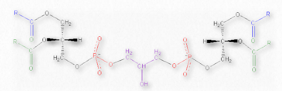
The molecules appear to be required for both Complex IV and Complex III of the electron transport chain to maintain its structure. The ATP synthase enzyme (Complex V) of the oxidative phosphorylation system also binds four molecules of cardiolipin. It has been proposed that cardiolipin functions as a proton trap in the process of proton pumping by Complex IV.

Cardiolipin also plays a role in apoptosis. As shown in Figure 2.203, oxidation of cardiolipin by a cardiolipin-specific oxygenase causes cardiolipin to move from the inner mitochondrial membrane to the outer one, helping to form a permeable pore and facilitating the transport of cytochrome c out of the intermembrane space and into the cytoplasm - a step in the process of apoptosis.Plasmalogens
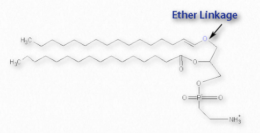
A special class of the glycerophospholipids are the plasmalogens (Figure 2.209). They differ in containing a vinyl ether linkage at position 1 of glycerol, in contrast to other glycerophopsholipids, which have an ester linkage at this position. Position 2 of each is an ester. The precursor for the ether linkage is typically a 16 or 18 carbon saturated alcohol or an 18 carbon unsaturated alcohol.
At the phosphate tail, the most commonly attached groups are ethanolamine or choline. Plasmalogens are found abundantly in humans in heart (30-40% of choline phospholipids). 30% of the glycerophospholipids in brain are plasmalogens and 70% of the ethanolamine lipids of the myelin sheath of nerve cells are plasmalogens.
Though their function is not understood, it is believed that plasmalogens may provide some protection against reactive oxygen species and have roles in signaling.
Lecithin
Lecithin is a generic term for a combination of lipid substances that includes phosphoric acid, glycerol, glycolipids, triglycerides, and phospholipids. Lecithin is a wetting agent helpful with emulsification and encapsulation and is even used as an anti-sludge additive in motor lubricants. Lecithin is used in candy bars to keep cocoa and cocoa butter from separating. Though considered safe as a food ingredient, lecithin can be converted by gut bacteria to trimethylamine-N-oxide which may contribute to cholesterol deposition and atherosclerosis.
Sphingolipids
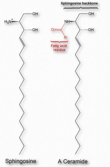
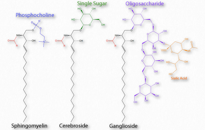
Fatty acids are also components of a broad class of molecules called sphingolipids. Sphingolipids are structurally similar to glycerophospholipids, though they are synthesized completely independently of them starting with palmitic acid and the amino acid serine. Sphingolipids are named for the amino alcohol known as sphingosine (Figure 2.210), though they are not directly synthesized from it. Figure 2.211 shows the generalized structure of sphingolipids.
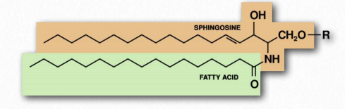
If the R-group is a hydrogen, the molecule is called a ceramide. When the R-group is phosphoethanolamine the resulting molecule is sphingomyelin, an important component of the myelin sheath and lipid membranes. If a single, simple sugar is instead added, a cerebroside is created (Figure 2.212). Addition of a complex oligosaccharide creates a ganglioside.
Complex sphingolipids may play roles in cellular recognition and signaling. Sphingolipids are found most abundantly in plasma membrane and are almost completely absent from mitochondrial and endoplasmic reticulum membranes. In animals, dietary sphingolipids have been linked to reduced colon cancer, reductions in LDLs, and increases in HDLs. Like the glycerophospholipids, sphingolipids are amphiphilic. Most sphingolipids except sphingomyelin do not contain phosphate.
Signaling lipids involved in inflammation
Eicosanoids
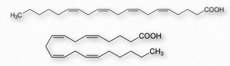
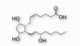
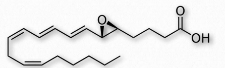
Fatty acids made from omega-6 and omega-3 fatty acids include three important fatty acids containing 20 carbons. They include arachidonic acid (an ω-6 fatty acid with four double bonds (Δ-5,8,11,14) - Figure 2.213), eicosapentaenoic acid (an ω-3 fatty acid with five double bonds, and dihomo-γ-linolenic acid (an ω-6 fatty acid with three double bonds). The class of compounds known as eicosanoids is made by oxidation of these compounds. Subclasses include include prostaglandins, prostacyclins, thromboxanes, lipoxins, leukotrienes, and endocannabinoids (Figures 2.214-2.219). Eicosanoids play important roles affecting inflammation, immunity, mood, and behavior.
Prostaglandins
A collection of molecules acting like hormones, prostaglandins are derived from arachidonic acid and have many differing (even conflicting) physiological effects. These include constriction or dilation of vascular smooth muscle cells, induction of labor, regulation of inflammation, and action on the thermoregulatory center of the hypothalamus to induce fever, among others.
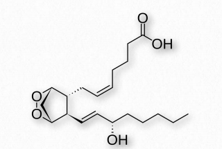
Prostaglandins are grouped with the thromboxanes (below) and prostacyclins (below), as prostanoids. The prostanoids, which all contain 20 carbons are a subclass of the eicosanoids. Prostaglandins are found in most tissues of higher organisms. They are autocrine or paracrine compounds produced from essential fatty acids. The primary precursor of the prostaglandins is the fatty acid known as arachidonic acid and the prostaglandin made from it is known as PGH2 (Figure 2.214), which, in turn is a precursor of other prostaglandins, as well as the prostacyclins and thromboxanes.
Interesting prostaglandins
PGD2 - inhibits hair follicle growth, vasodilator, causes bronchial constriction, higher in lungs of asthmatics than others.
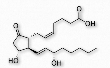
PGE2 (Figure 2.215) - exerts effects in labor (soften cervix, uterine contraction), stimulates bone resorption by osteoclasts, induces fever, suppresses T-cell receptor signaling, vasodilator, inhibits release of noradrenalin from sympathetic nerve terminals. It is a potent activator of the Wnt signaling pathway.
A prostaglandin can have opposite effects, depending on which receptor it binds to. Binding of PGE2 to the EP1 receptor causes bronchoconstriction and smooth muscle contraction, whereas binding of the same molecule to the EP2 receptor causes bronchodilation and smooth muscle relaxation.
PGF2α (Figure 2.216)- uterine contractions, induces labor, bronchoconstriction.
PGI2 - vasodilation, bronchodilation, inhibition of platelet aggregation.
Thromboxanes
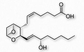
Thromboxanes play roles in clot formation and named for their role in thrombosis. They are potent vasoconstrictors and facilitate platelet aggregation. They are synthesized in platelets, as well. The anti-clotting effects of aspirin have their roots in the inhibition of synthesis of PGH2, which is the precursor of the thromboxanes. The most common thromboxanes are A2 (Figure 2.217) and B2.
Prostacyclin
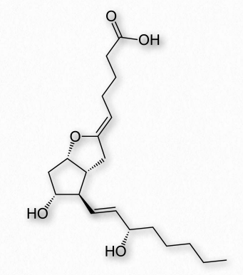
Prostacyclin (also known as prostaglandin I2 or PGI2 - Figure 2.218) counters the effects of thromboxanes, inhibiting platelet activation and acting as vasodilators. It is produced from PGH2 by action of the enzyme prostacyclin synthase.
Leukotrienes
Another group of eicosanoid compounds are the leukotrienes (Figure 2.219). Like prostaglandins, leukotrienes are made from arachidonic acid. The enzyme catalyzing their formation is a dioxygenase known as arachidonate 5-lipoxygenase. Leukotrienes are involved in regulating immune responses. They are found in leukocytes and other immunocompetent cells, such as neutrophils, monocytes, mast cells, eosinophils, and basophils. Leukotrienes are associated with production of histamines and prostaglandins, which act as mediators of inflammation. Leukotrienes also trigger contractions in the smooth muscles of the bronchioles. When overproduced, they may pay a role in asthma and allergic reactions. Some treatments for asthma aim at inhibiting production or action of leukotrienes.
Lipid-soluble vitamins are critical for enzyme function
Vitamin A
Vitamin A comes in three primary chemical forms, retinol (storage in liver - Figure 2.225), retinal (role in vision - Figure 2.226), and retinoic acid (roles in growth and development). All vitamin A forms are diterpenoids and differ only in the chemical form of the terminal group. Retinol is mostly used as the storage form of the vitamin.
Retinol is commonly esterified to a fatty acid and kept in the liver. In high levels, the compound is toxic. Retinol is obtained in the body by hydrolysis of the ester or by reduction of retinal. Importantly, neither retinal nor retinol can be made from retinoic acid. Retinoic acid is important for healthy skin and teeth, as well as bone growth. It acts in differentiation of stem cells through a specific cellular retinoic acid receptor.
Sources
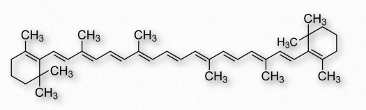
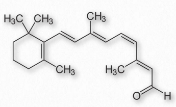
Good sources of vitamin A are liver and eggs, as well as many plants, including carrots, which have a precursor, β-carotene (Figure 2.227) from which retinol may be made by action of a dioxygenase.
Light sensitivity The conjugated double bond system in the side chain of vitamin A is sensitive to light and can flip between cis and trans forms on exposure to it. It is this response to light that makes it possible for retinal to have a role in vision in the rods and cones of the eyes. Here, the aldehyde form (retinal) is bound to the protein rhodopsin in the membranes of rod and cone cells.
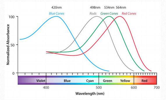
When exposed to light of a particular wavelength, the “tail” of the retinal molecule will flip back and forth from cis to trans at the double bond at position 11 of the molecule. When this happens, a nerve signal is generated that signals the brain of exposure to light. Slightly different forms of rhodopsin have different maximum absorption maxima allowing the brain to perceive red, green and blue specifically and to assemble those into the images we see (Figure 2.228). Cones are the cells responsible for color vision, whereas rods are mostly involved in light detection in low light circumstances.
Deficiency and surplus
Deficiency of vitamin A is common in developing countries and was inspiration for the design and synthesis of the geneticallymodified golden rice, which is used as a source of vitamin A to help prevent blindness in children. Overdose of vitamin A, called hypervitaminosis A is dangerous and can be fatal. Excess vitamin A is also suspected to be linked to osteoporosis. In smokers, excess vitamin A is linked to an increased rate of lung cancer, but non-smokers have a reduced rate.
Vitamin D
The active form of vitamin D plays important roles in the intestinal absorption of calcium and phosphate and thus in healthy bones. Technically, vitamin D isn’t even a vitamin, as it is a compound made by the body. Rather, it behaves more like a hormone.
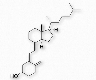
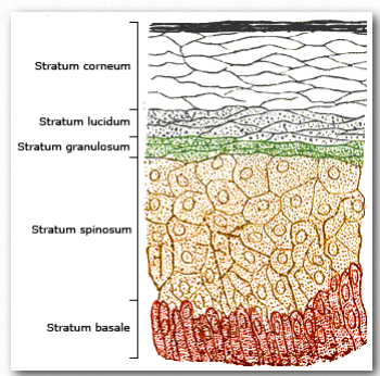
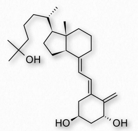
Derived from ultimately from cholesterol, vitamin D can be made in a reaction catalyzed by ultraviolet light. In the reaction, the intermediate 7-dehydrocholesterol is converted to cholecalciferol (vitamin D3) by the uv light (Figure 2.229). The reaction occurs most readily in the bottom two layers of the skin shown in Figure 2.230.
Forms of vitamin D
Five different compounds are referred to as vitamin D. They are
Vitamin D1 - A mixture of ergocalciferol and lumisterol
Vitamin D2 - Ergocalciferol
Vitamin D3 - Cholecalciferol Vitamin
D4 - 22-Dihydroergocalciferol Vitamin
D5 - Sitocalciferol
Vitamin D3 is the most common form used in vitamin supplements and it and vitamin D2 are commonly obtained in the diet, as well. The active form of vitamin D, calcitriol (Figure 2.231), is made in the body in controlled amounts. This proceeds through two steps from cholecalciferol. First, a hydroxylation in the liver produces calcidiol and a second hydroxylation in the kidney produces calcitriol. Monocyte macrophages can also synthesize vitamin D and they use is as a cytokine to stimulate the innate immune system.
Mechanism of action
Calcitriol moves in the body bound to a vitamin D binding protein, which delivers it to target organs. Calcitriol inside of cells acts by binding a vitamin D receptor (VDR), which results in most of the vitamin’s physiological effects. After binding calcitriol, the VDR migrates to the nucleus where it acts as a transcription factor to control levels of expression of calcium transport proteins (for example) in the intestine. Most tissues respond to VDR bound to calcitriol and the result is moderation of calcium and phosphate levels in cells.
Deficiency/excess
Deficiency of vitamin D is a cause of the disease known as rickets, which is characterized by soft, weak bones and most commonly is found in children. It is not common in the developed world, but elsewhere is of increasing concern.
Excess of vitamin D is rare, but has toxic effects, including hypercalcemia, which results in painful calcium deposits in major organs. Indications of vitamin D toxicity are increased urination and thirst. Vitamin D toxicity can lead to mental retardation and many other serious health problems.
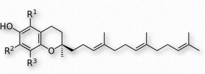
Vitamin E
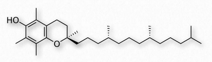
Vitamin E comprises a group of two compounds (tocopherols and tocotrienols - Figure 2.232) and stereoisomers of each. It is commonly found in plant oils. The compounds act in cells as fat-soluble antioxidants. α-tocopherol (Figure 2.233), the most active form of the vitamin, works 1) through the glutathione peroxidase protective system and 2) in membranes to interrupt lipid peroxidation chain reactions. In both actions, vitamin E reduces levels of reactive oxygen species in cells.
Action
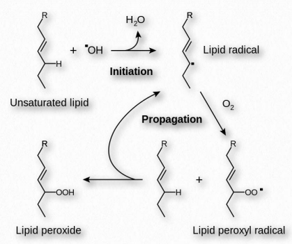
Vitamin E scavenges oxygen radicals (possessing unpaired electrons) by reacting with them to produce a tocopheryl radical. This vitamin E radical can be converted back to its original form by a hydrogen donor. Vitamin C is one such donor. Acting in this way, Vitamin E helps reduce oxidation of easily oxidized compounds in the lipid peroxidation reactions (Figure 2.234).
Vitamin E also can affect enzyme activity. The compound can inhibit action of protein kinase C in smooth muscle and simultaneously activate catalysis of protein phosphatase 2A to remove phosphates, stopping smooth muscle growth.
Deficiency/excess
Deficiency of vitamin E can lead to poor conduction of nerve signals and other issues arising from nerve problems. Low levels of the vitamin may be a factor in low birth weights and premature deliveries. Deficiency, however, is rare, and not usually associated with diet.
Excess Vitamin E reduces vitamin K levels, thus reducing the ability to clot blood. Hypervitaminosis of vitamin E in conjunction with aspirin can be life threatening. At lower levels, vitamin E may serve a preventative role with respect to atherosclerosis by reducing oxidation of LDLs, a step in plaque formation.
Vitamin K
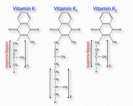
Like the other fat-soluble vitamins, Vitamin K comes in multiple forms (Figure 2.235) and is stored in fat tissue in the body. There are two primary forms of the vitamin - K1 and K2 and the latter has multiple sub-forms . Vitamins K3, K4, and K5 are made synthetically, not biologically.
Action
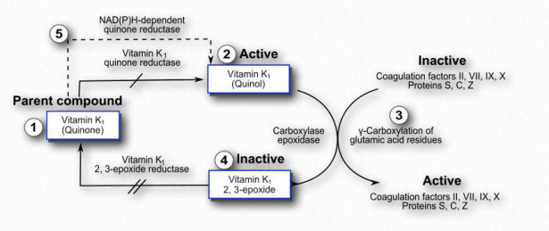
Vitamin K is used as a co-factor for enzymes that add carboxyl groups to glutamate side chains of proteins to increase their affinity for calcium. Sixteen such proteins are known in humans. They include proteins involved in blood clotting (prothrombin (called Factor II), Factors VII, IX, and X), bone metabolism (osteocalcin, also called bone Gla protein (BGP), matrix Gla protein (MGP), and periostin) and others.
Modification of prothrombin is an important step in the process of blood clotting (see HERE). Reduced levels of vitamin K result in less blood clotting, a phenomenon sometimes referred to as blood thinning. Drugs that block recycling of vitamin K (Figure 2.236) by inhibiting the vitamin K epoxide reductase, produce lower levels of the vitamin and are employed in treatments for people prone to excessive clotting. Warfarin (coumadin) is one such compound that acts in this way and is used therapeutically. Individuals respond to the drug differentially, requiring them to periodically be tested for levels of clotting they possess, lest too much or too little occur.
Sources
Vitamin K1 is a stereoisomer of the plant photosystem I electron receptor known as phylloquinone and is found abundantly in green, leafy vegetables. Phylloquinone is one source of vitamin K, but the compound binds tightly to thylakoid membranes and tends to have low bioavailability. Vitamin K2 is produced by microbes in the gut and is a primary source of the vitamin. Infants in the first few days before they establish their gut flora and people taking broad spectrum antibiotics may have reduced levels, as a result. Dietary deficiency is rare in the absence of damage to the small bowel. Others at risk of deficiency include people with chronic kidney disease and anyone suffering from a vitamin D deficiency. Deficiencies produce symptoms of easy bruising, heavy menstrual bleeding, anemia, and nosebleeds.
Other lipids
Cannabinoids
Cannabinoids are a group of chemicals that bind to and have effects on brain receptors (cannabinoid receptors), repressing neurotransmitter release. Classes of these compounds include endocannabinoids (made in the body), phytocannabinoids (made in plants, such as marijuana), and synthetic cannabinoids (man-made).
Endocannabinoids are natural molecules derived from arachidonic acid. Cannabinoid receptors are very abundant, comprising the largest number of G-protein- 247 Figure 2.243 - Tetrahydrocannabinol - Active ingredient in marijuana coupled receptors found in brain. The best known phytocannabinoid is Δ-9- tetrahydrocannabinol (THC), the primary psychoactive ingredient (of the 85 cannabinoids) of marijuana (Figure 2.243).

Anandamide
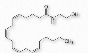
Anandamide (N-arachidonoylethanolamine - Figure 2.244) is an endocannabinoid neurotransmitter derived from arachidonic acid. It exerts its actions primarily through the CB1 and CB2 cannabinoid receptors, the same ones bound by the active ingredient in marijuana, Δ9-tetrahydrocannabinol. Anandamide has roles in stimulating eating/appetite and affecting motivation and pleasure. It has been proposed to play a role in “runners high,” an analgesic effect experienced from exertion, especially among runners. Anandamide appears to impair memory function in rats.
Anandamide has been found in chocolate and two compounds that mimic its effects (N-oleoylethanolamine and Nlinoleoylethanolamine) are present as well. The enzyme fatty acid amide hydrolase (FAAH) breaks down anandamide into free arachidonic acid and ethanolamine.
Lipoxins
Lipoxins (Figure 2.245) are eicosanoid compounds involved in modulating immune responses and they have anti-inflammatory effects. When lipoxins appear in inflammation it begins the end of the process. Lipoxins act to attract macrophages to apoptotic cells at the site of inflammation and they are engulfed. Lipoxins further act to start the resolution phase of the inflammation process.
At least one lipoxin (aspirin-triggered LX4) has its synthesis stimulated by aspirin. This occurs as a byproduct of aspirin’s acetylation of COX-2. When this occurs, the enzyme’s catalytic activity is re-directed to synthesis of 15R-hydroxyeicosatetraenoic acid (HETE) instead of prostaglandins. 15R-HETE is a procursor of 15-epimer lipoxins, including aspirin-triggered LX4.
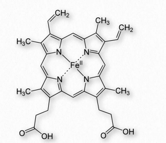
Heme
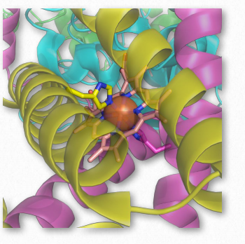
Heme groups are a collection of protein/ enzyme cofactors containing a large heterocyclic aromatic ring known as a porphyrin ring with a ferrous (Fe++) ion in the middle. An example porphyrin ring with an iron (found in Heme B of hemoglobin), is shown in Figure 2.246. When contained in a protein, these are known collectively as hemoproteins (Figure 2.247).
Heme, of course, is a primary component of hemoglobin, but it is also found in other proteins, such as myoglobin, cytochromes, and the enzymes catalase and succinate dehydrogenase. Hemoproteins function in oxygen transport, catalysis, and electron transport. Heme is synthesized in the liver and bone marrow in a pathway that is conserved across a wide range of biology.
Porphobilinogen
Porphobilinogen (Figure 2.248) is a pyrrole molecule involved in porphyrin metabolism. It is produced from aminolevulinate by action of the enzyme known as ALA dehydratase. Porphobilinogen is acted upon by the enzyme porphobilinogen deaminase. Deficiency of the latter enzyme (and others in porphyrin metabolism) can result in a condition known as porphyria, which results in accumulation of porphobilinogen in the cytoplasm of cells.
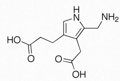
The disease can manifest itself with acute abdominal pain and numerous psychiatric issues. Both Vincent van Gogh and King ` George III are suspected to have suffered from porphyria, perhaps causing the “madness of King George III.” Porphyria is also considered by some to be the impetus for the legend of vampires seeking blood from victims, since the color of the skin in non-acute forms of the disease can be miscolored, leading some to perceive that as a deficiency of hemoglobin and hence the “thirst” for blood imagined for vampires.
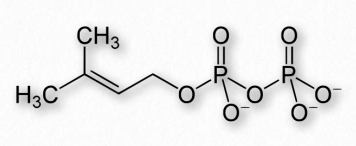
Dolichols
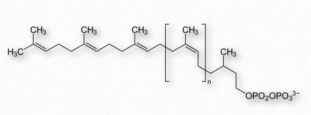
Dolichol is a name for a group of non-polar molecules made by combining isoprene units together. Phosphorylated forms of dolichols play central roles in the N-glycosylation of proteins. This process, which occurs in the endoplasmic reticulum of eukaryotic cells, begins with a membrane-embedded dolichol pyrophosphate (Figure 2.249) to which an oligosaccharide is attached (also see HERE). This oligosaccharide contains three molecules of glucose, nine molecules of mannose and two molecules of N-acetylglucosamine.
Interestingly, the sugars are attached to the dolichol pyrophosphate with the pyrophosphate pointing outwards (away from) the endoplasmic reticulum, but after attachment, the dolichol complex flips so that the sugar portion is situated on the inside of the endoplasmic reticulum. There, the entire sugar complex is transferred to the amide of an asparagine side chain of a target protein.
The only asparagine side chains to which the attachment can be made are in proteins where the sequences Asn-X-Ser or Asn-X-Thr occur. Sugars can be removed/added after the transfer to the protein. Levels of dolichol in the human brain increase with age, but in neurodegenerative diseases, they decrease.
Terpenes
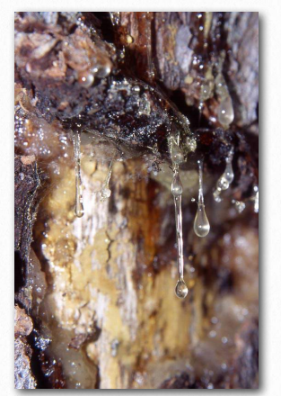
Terpenes are members of a class of nonpolar molecules made from isoprene units. Terpenes are produced primarily by plants and by some insects. Terpenoids are a related group of molecules that contain functional groups lacking in terpenes.
Terpenes have a variety of functions. In plants, they often play a defensive role protecting from insects. The name of terpene comes from turpentine, which has an odor like some of the terpenes. Terpenes are common components of plant resins (think pine) and they are widely used in medicines and as fragrances. Hops, for example, gain some of their distinctive aroma and flavor from terpenes. Not all terpenes, however have significant odor.
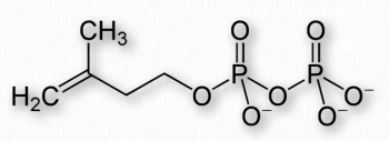
Synthesis
Terpenes, like steroids, are synthesized starting with simple building blocks known as isoprenes. There are two of them - dimethylallyl pyrophosphate and the related isopentenyl pyrophosphate and (Figures 2.252 and 2.253) which combine 1-2 units at a time to make higher order structures. Terpene synthesis overlaps and includes steroid synthesis.
Terpenes and terpenoids are classified according to how many isoprene units they contain. They include hemiterpenes (one unit), monoterpenes (two units), sesquiterpenes (three units), diterpenes (four units), sesterterpenes (five units), triterpenes (six units), sesquarterpenes (seven units), tetraterpenes (eight units), polyterpenes (many units). Another class of terpene-containing molecules, the norisoterpenoids arise from peroxidase-catalyzed reactions on terpene molecules.
Examples
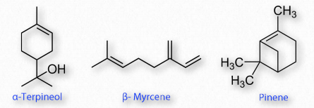
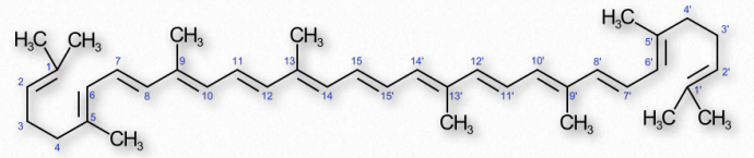
Common terpenes include monoterpenes of terpineol (lilacs), limonene (citrus), myrcene (hops), linalool (lavender), and pinene (pine). Higher order terpenes include taxadiene (diterpene precursor of taxol), lycopene (tetraterpenes), carotenes (tetraterpenes), and natural rubber (polyterpenes).
Steroid precursors geranyl pyrophosphate (monoterpene derivative), farnesyl pyrophosphate (sesquiterpene derivative), and squalene (triterpene) are all terpenes or derivatives of them. Vitamin A and phytol are derived from diterpenes.
Caffeine
Caffeine is the world’s most actively consumed psychoactive drug (Figure 2.255). A methylxanthine alkaloid, caffeine is closely related to adenine and guanine and this is responsible for many effects on the body. Caffeine blocks the binding of adenosine on its receptor and consequently prevents the onset of drowsiness induced by adenosine. Caffeine readily crosses the blood-brain barrier and stimulates release of neurotransmitters. Caffeine stimulates portions of the autonomic nervous system and inhibits the activity of phosphodiesterase. The latter has the result of raising cAMP levels in cells, which activates protein kinase A and activates glycogen breakdown, inhibits TNF-α and leukotriene synthesis, which results in reduction of inflammation and innate immunity.
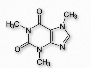
Caffeine also has effects on the cholinergic system (acetylcholinesterase inhibitor), is an inositol triphosphate receptor 1 antagonist, and is a voltage independent activator of ryanodin receptors (a group of calcium channels found in skeletal muscle, smooth muscle, and heart muscle cells).
The half-life of caffeine in the body varies considerably. In healthy adults, it has a half-life of about 3-7 hours. Nicotine decreases the half-life and contraceptives and pregnancy can double it. The liver metabolizes caffeine, so the health of the liver is a factor in the halflife. CYP1A2 of the cytochrome P450 oxidase enzyme is primarily responsible. Caffeine is a natural pesticide in plants, paralyzing predator bugs.