Radiation in Biology and Medicine
- Page ID
- 1476
\( \newcommand{\vecs}[1]{\overset { \scriptstyle \rightharpoonup} {\mathbf{#1}} } \)
\( \newcommand{\vecd}[1]{\overset{-\!-\!\rightharpoonup}{\vphantom{a}\smash {#1}}} \)
\( \newcommand{\id}{\mathrm{id}}\) \( \newcommand{\Span}{\mathrm{span}}\)
( \newcommand{\kernel}{\mathrm{null}\,}\) \( \newcommand{\range}{\mathrm{range}\,}\)
\( \newcommand{\RealPart}{\mathrm{Re}}\) \( \newcommand{\ImaginaryPart}{\mathrm{Im}}\)
\( \newcommand{\Argument}{\mathrm{Arg}}\) \( \newcommand{\norm}[1]{\| #1 \|}\)
\( \newcommand{\inner}[2]{\langle #1, #2 \rangle}\)
\( \newcommand{\Span}{\mathrm{span}}\)
\( \newcommand{\id}{\mathrm{id}}\)
\( \newcommand{\Span}{\mathrm{span}}\)
\( \newcommand{\kernel}{\mathrm{null}\,}\)
\( \newcommand{\range}{\mathrm{range}\,}\)
\( \newcommand{\RealPart}{\mathrm{Re}}\)
\( \newcommand{\ImaginaryPart}{\mathrm{Im}}\)
\( \newcommand{\Argument}{\mathrm{Arg}}\)
\( \newcommand{\norm}[1]{\| #1 \|}\)
\( \newcommand{\inner}[2]{\langle #1, #2 \rangle}\)
\( \newcommand{\Span}{\mathrm{span}}\) \( \newcommand{\AA}{\unicode[.8,0]{x212B}}\)
\( \newcommand{\vectorA}[1]{\vec{#1}} % arrow\)
\( \newcommand{\vectorAt}[1]{\vec{\text{#1}}} % arrow\)
\( \newcommand{\vectorB}[1]{\overset { \scriptstyle \rightharpoonup} {\mathbf{#1}} } \)
\( \newcommand{\vectorC}[1]{\textbf{#1}} \)
\( \newcommand{\vectorD}[1]{\overrightarrow{#1}} \)
\( \newcommand{\vectorDt}[1]{\overrightarrow{\text{#1}}} \)
\( \newcommand{\vectE}[1]{\overset{-\!-\!\rightharpoonup}{\vphantom{a}\smash{\mathbf {#1}}}} \)
\( \newcommand{\vecs}[1]{\overset { \scriptstyle \rightharpoonup} {\mathbf{#1}} } \)
\( \newcommand{\vecd}[1]{\overset{-\!-\!\rightharpoonup}{\vphantom{a}\smash {#1}}} \)
\(\newcommand{\avec}{\mathbf a}\) \(\newcommand{\bvec}{\mathbf b}\) \(\newcommand{\cvec}{\mathbf c}\) \(\newcommand{\dvec}{\mathbf d}\) \(\newcommand{\dtil}{\widetilde{\mathbf d}}\) \(\newcommand{\evec}{\mathbf e}\) \(\newcommand{\fvec}{\mathbf f}\) \(\newcommand{\nvec}{\mathbf n}\) \(\newcommand{\pvec}{\mathbf p}\) \(\newcommand{\qvec}{\mathbf q}\) \(\newcommand{\svec}{\mathbf s}\) \(\newcommand{\tvec}{\mathbf t}\) \(\newcommand{\uvec}{\mathbf u}\) \(\newcommand{\vvec}{\mathbf v}\) \(\newcommand{\wvec}{\mathbf w}\) \(\newcommand{\xvec}{\mathbf x}\) \(\newcommand{\yvec}{\mathbf y}\) \(\newcommand{\zvec}{\mathbf z}\) \(\newcommand{\rvec}{\mathbf r}\) \(\newcommand{\mvec}{\mathbf m}\) \(\newcommand{\zerovec}{\mathbf 0}\) \(\newcommand{\onevec}{\mathbf 1}\) \(\newcommand{\real}{\mathbb R}\) \(\newcommand{\twovec}[2]{\left[\begin{array}{r}#1 \\ #2 \end{array}\right]}\) \(\newcommand{\ctwovec}[2]{\left[\begin{array}{c}#1 \\ #2 \end{array}\right]}\) \(\newcommand{\threevec}[3]{\left[\begin{array}{r}#1 \\ #2 \\ #3 \end{array}\right]}\) \(\newcommand{\cthreevec}[3]{\left[\begin{array}{c}#1 \\ #2 \\ #3 \end{array}\right]}\) \(\newcommand{\fourvec}[4]{\left[\begin{array}{r}#1 \\ #2 \\ #3 \\ #4 \end{array}\right]}\) \(\newcommand{\cfourvec}[4]{\left[\begin{array}{c}#1 \\ #2 \\ #3 \\ #4 \end{array}\right]}\) \(\newcommand{\fivevec}[5]{\left[\begin{array}{r}#1 \\ #2 \\ #3 \\ #4 \\ #5 \\ \end{array}\right]}\) \(\newcommand{\cfivevec}[5]{\left[\begin{array}{c}#1 \\ #2 \\ #3 \\ #4 \\ #5 \\ \end{array}\right]}\) \(\newcommand{\mattwo}[4]{\left[\begin{array}{rr}#1 \amp #2 \\ #3 \amp #4 \\ \end{array}\right]}\) \(\newcommand{\laspan}[1]{\text{Span}\{#1\}}\) \(\newcommand{\bcal}{\cal B}\) \(\newcommand{\ccal}{\cal C}\) \(\newcommand{\scal}{\cal S}\) \(\newcommand{\wcal}{\cal W}\) \(\newcommand{\ecal}{\cal E}\) \(\newcommand{\coords}[2]{\left\{#1\right\}_{#2}}\) \(\newcommand{\gray}[1]{\color{gray}{#1}}\) \(\newcommand{\lgray}[1]{\color{lightgray}{#1}}\) \(\newcommand{\rank}{\operatorname{rank}}\) \(\newcommand{\row}{\text{Row}}\) \(\newcommand{\col}{\text{Col}}\) \(\renewcommand{\row}{\text{Row}}\) \(\newcommand{\nul}{\text{Nul}}\) \(\newcommand{\var}{\text{Var}}\) \(\newcommand{\corr}{\text{corr}}\) \(\newcommand{\len}[1]{\left|#1\right|}\) \(\newcommand{\bbar}{\overline{\bvec}}\) \(\newcommand{\bhat}{\widehat{\bvec}}\) \(\newcommand{\bperp}{\bvec^\perp}\) \(\newcommand{\xhat}{\widehat{\xvec}}\) \(\newcommand{\vhat}{\widehat{\vvec}}\) \(\newcommand{\uhat}{\widehat{\uvec}}\) \(\newcommand{\what}{\widehat{\wvec}}\) \(\newcommand{\Sighat}{\widehat{\Sigma}}\) \(\newcommand{\lt}{<}\) \(\newcommand{\gt}{>}\) \(\newcommand{\amp}{&}\) \(\definecolor{fillinmathshade}{gray}{0.9}\)What comes to mind when you think of radiation? If you ask the average American, radiation would conjure images of deformed humans and malignant diseases, namely cancer. Despite radiation's notorious history, radiation has revolutionized the medical field, enhancing the ability of medical professionals to treat and diagnose diseases.
Introduction
Imagine a patient enters the hospital complaining of severe chest pain. After consulting the patient, the doctors decide that the source of the patient's problems is a constricted artery. However, the human body contains several long arteries. The doctors could use surgery to find the source of the clot, but this could take many hours and with no definite guarantee of success.This is exactly where radiation helps medical professionals bridge the gap that once existed. Now, CT scans, short for computed tomography, use X-ray particles along with advanced computer technology in order to produce highly detailed cross sectional images of the body. Using these scans, medical professionals can accurately find clots and other other harmful medical conditions.
In PET scans, short for positron imagery tomography, tracers are injected into the brain. These tracers emit gamma rays, which are picked up by a computer which use the gamma radiation in order to construct a two dimensional image of the brain. With the radiation coupled with an advanced computer, a two dimensional image is displayed, providing medical professionals with a view to the metabolic activity of the brain. On top of assisting medical professionals with diagnosing diseases, radiation also has therapeutic powers.
Radiation Therapy is used as a treatment to control malignant cells within cancer patients. Oncologists (specialists that deals with cancer) used radiation therapy frequently to help slow or cure the spread of cancer within inidivduals. Radiation is specifically applied to malignant tumors in order to shrink them in size, while hopefully not causing more damage to surrounding healthy cells. Medical professionals, mainly radiation oncologists, administer a variety of dosages to patient, contingent to the patients current health, as well as other treatments such as chemotherapy, success of surgery, etc.
Radiation in Imaging
X-Ray Imaging
German physicist, Wilhem Röntgen, discovered eletromagnetic radiation on 8 November 1895. In addition to Röntgen receiving the first Nobel Prize in Physics (1901), the use of the x-ray has become instrumental in modern medicine. Lastly, because of his discovery Röntgen is considered the father of diagnositic radiology.
X-Ray imaging is one of the most basic and routine forms of imaging within modern medicine. Using electromagnetic radiation, scientists and doctors have the ability to visualize internal situations. X-Ray imaging takes advantage of the fact that dense structures (i.e. bone) absorb more x-rays than the softer tissues that surround it. By projecting radiation onto special film paper, the denser substance casts a shadow where less x-rays pass through the body, and a contrasting image is produced. The radiation emitted by an X-Ray is of the wavelength of 10 to 0.01 nanometers(nm). Because some parts of the body may be thicker than others, X-Rays are used in "soft" (10 to 0.10 nm) and "hard" (0.10 to 0.01 nm) dosages or radiation.
X-rays are produced by passing an electron beam through a tungsten anode in a vacuum. The electromagnetic waves that are released can be focused and adjusted according to what is imaged. X-Rays can image in very high definition at a low cost, but they provide poor contrast in soft tissues. Contrast agents can also be added to the body prior to imaging, which allows different tissues to absorb more radiation, creating a better contrasting image. This allows doctors and scientists to work with much more specific images within their diagnosis or research (i.e. the ability to determine different aspects of soft-tissue).
CT Scanning
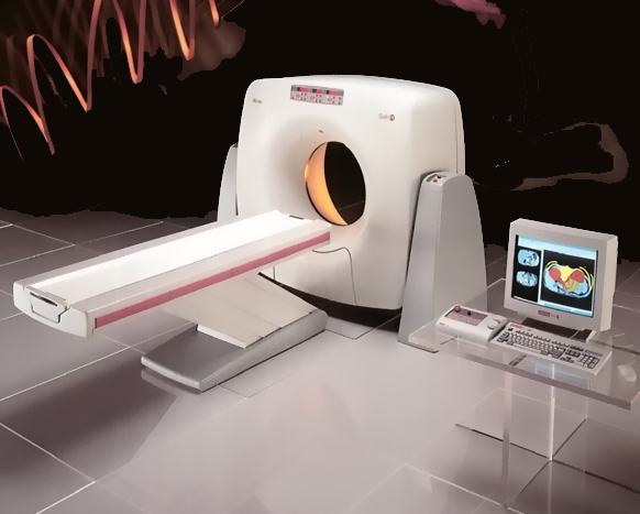
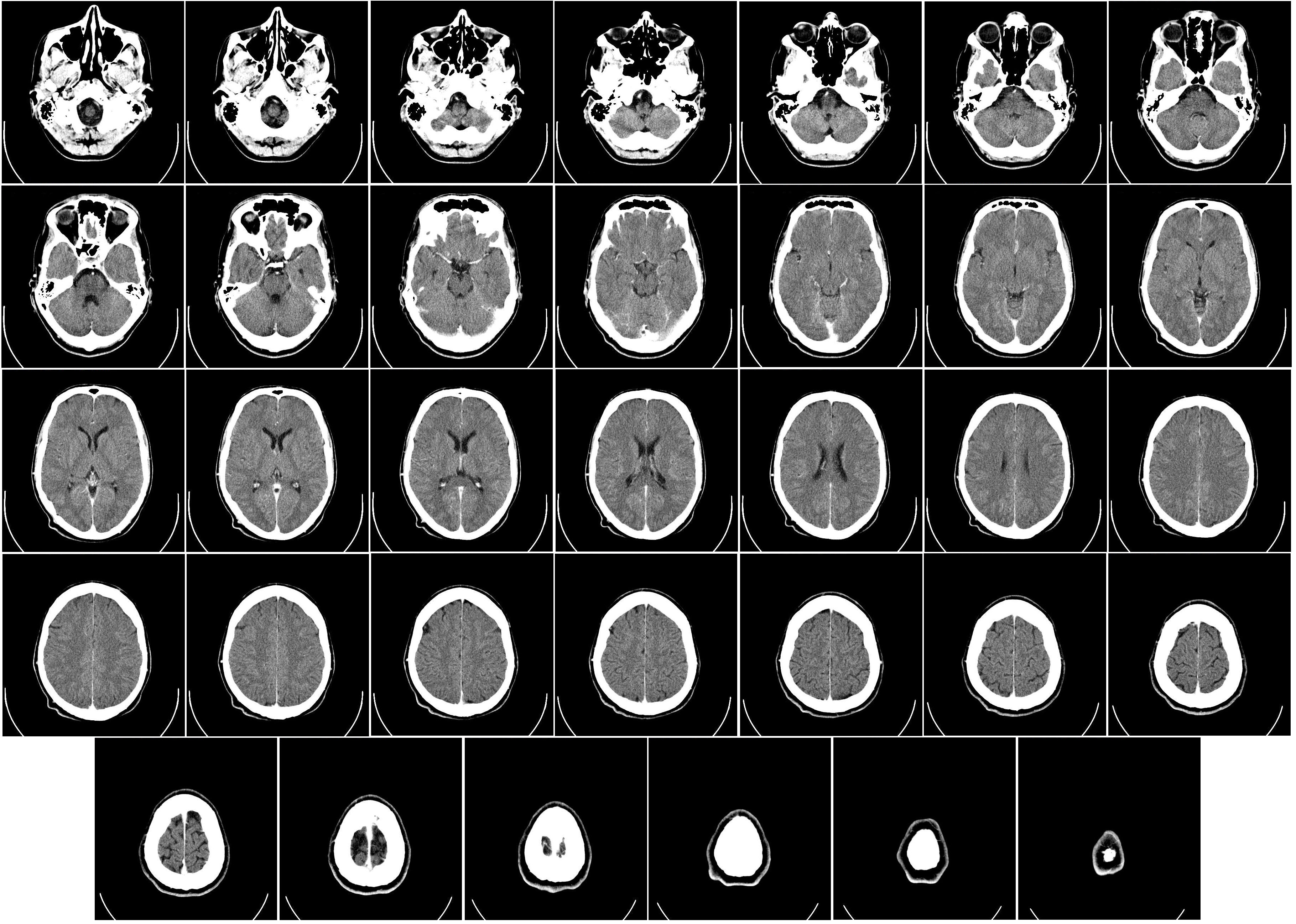
CT/ CAT (Computerized Axial Tomography) scans have the same fundamentals of x-rays, but they produce 3-D images using tomography. Using digitial geometric processing, the CT scan creates a 3-D image compiling a large number of 2-D x-rays. These 2-D images are thin cross sections of the body taken from a single axis of rotation. A CT scanner consists of a table where the patient lies on, an x-ray device which rotates at high speed around the patient taking hundreds of cross sections of the body, and a computer with appropriate software to compile and render the 3-D image. The table in Figure 1 moves through the tube, allowing the machine to take hundreds of pictures from head to toe.
Just like with x-ray, contrast agents are typically added to the body before imaging occurs so that different tissues are more defined. Due to the fact that many more images are being taken in order to form a complete three dimensional image, the radiation dose is much higher. While a chest x-ray is around .1 mSv or radiation, a chest CAT scan subjects the patient to about 8 mSv of radiation; over twice the amount that the body would absorb from its environment in a year. For this reason, it is uncommon and unsafe to have multiple CAT scans taken unless completely necessary.

Ironically, while X-Rays are used for medicine, they do pose health risks upon patients, especially CT Scans. Stated by the International Agenct for Research on Cancer, X-Ray radiation is considered a carcinogen (cancer causing). Measured in Sievert(s) (Sv) or Gray(s) (Gy), are the units of how much radiation energy is desposited within the body. For example a chest X-Ray has a mSv of 0.1. In contrast, the average American will absorb 3 mSv of background radiation in a year. Despite the X-Ray of a chest posing a small amount or radiation in contrast with our annual intake of background radiation, there exist risks involved with radiation, especially of reproductive organs and the brain.

PET/SPECT Scanning
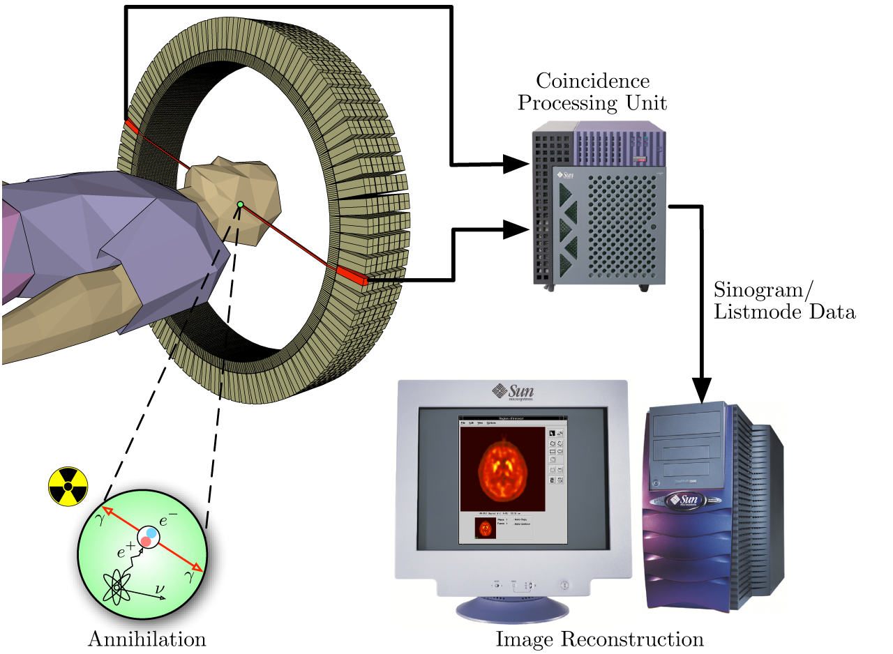
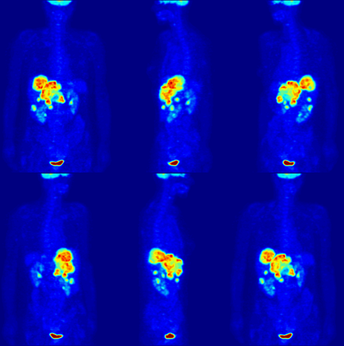
PET/ SPECT (Positron Emission Tomograpy/ Single Photon Emission Computerized Tomography) uses radioactively tagged molecules to image the body. These radioactive "radionuclides" give off gamma radiation, which eventually annihilates an electron. These radionuclides are isotopes with short half-lives, usually less than 30 minutes. This annihilation sends two gamma rays in exactly opposite directions with energy of 511keVs, which is picked up by a 5,000 to 20,000 gamma detectors that surround the patient. When two detectors on opposite sides of the patients body absorb gamma rays at the same time, the computer marks where the substance is and using this data can render a 3-D image (Figure 4).

Figure 3: upload.wikimedia.org/Wikipedia/commons/c/c1/PET-schema.png
PET Scanning, is used to image the physiological aspects of the body rather than the anatomy. It images the function of the body rather than the form, such as where tagged molecules go and how they are used. For instance, if you were to image the brain of a deceased person, nothing would show up on a PET scan opposed to a CAT scan, as the brain is no longer functional. Pet Scanning is very useful in imaging tumors, which can be done when patients are injected with certain tracers. Often times PET scanners are used in collaboration with CAT scanners to create a composite image that shows both the function and form of the body.

Figure 4:upload.wikimedia.org/Wikipedia/commons/3/3d/PET-MIPS-anim.gif
References
- Petrucci, Ralph H. General Chemistry. 9th ed. Upper Saddle River: Prentice Hall, 2007. Print
Contributors and Attributions
- Mike Reed (UC Davis), Eugene Kwon (UC Davis)

