12.3: Cells - Basic Units of Life
- Page ID
- 285366
\( \newcommand{\vecs}[1]{\overset { \scriptstyle \rightharpoonup} {\mathbf{#1}} } \)
\( \newcommand{\vecd}[1]{\overset{-\!-\!\rightharpoonup}{\vphantom{a}\smash {#1}}} \)
\( \newcommand{\id}{\mathrm{id}}\) \( \newcommand{\Span}{\mathrm{span}}\)
( \newcommand{\kernel}{\mathrm{null}\,}\) \( \newcommand{\range}{\mathrm{range}\,}\)
\( \newcommand{\RealPart}{\mathrm{Re}}\) \( \newcommand{\ImaginaryPart}{\mathrm{Im}}\)
\( \newcommand{\Argument}{\mathrm{Arg}}\) \( \newcommand{\norm}[1]{\| #1 \|}\)
\( \newcommand{\inner}[2]{\langle #1, #2 \rangle}\)
\( \newcommand{\Span}{\mathrm{span}}\)
\( \newcommand{\id}{\mathrm{id}}\)
\( \newcommand{\Span}{\mathrm{span}}\)
\( \newcommand{\kernel}{\mathrm{null}\,}\)
\( \newcommand{\range}{\mathrm{range}\,}\)
\( \newcommand{\RealPart}{\mathrm{Re}}\)
\( \newcommand{\ImaginaryPart}{\mathrm{Im}}\)
\( \newcommand{\Argument}{\mathrm{Arg}}\)
\( \newcommand{\norm}[1]{\| #1 \|}\)
\( \newcommand{\inner}[2]{\langle #1, #2 \rangle}\)
\( \newcommand{\Span}{\mathrm{span}}\) \( \newcommand{\AA}{\unicode[.8,0]{x212B}}\)
\( \newcommand{\vectorA}[1]{\vec{#1}} % arrow\)
\( \newcommand{\vectorAt}[1]{\vec{\text{#1}}} % arrow\)
\( \newcommand{\vectorB}[1]{\overset { \scriptstyle \rightharpoonup} {\mathbf{#1}} } \)
\( \newcommand{\vectorC}[1]{\textbf{#1}} \)
\( \newcommand{\vectorD}[1]{\overrightarrow{#1}} \)
\( \newcommand{\vectorDt}[1]{\overrightarrow{\text{#1}}} \)
\( \newcommand{\vectE}[1]{\overset{-\!-\!\rightharpoonup}{\vphantom{a}\smash{\mathbf {#1}}}} \)
\( \newcommand{\vecs}[1]{\overset { \scriptstyle \rightharpoonup} {\mathbf{#1}} } \)
\( \newcommand{\vecd}[1]{\overset{-\!-\!\rightharpoonup}{\vphantom{a}\smash {#1}}} \)
\(\newcommand{\avec}{\mathbf a}\) \(\newcommand{\bvec}{\mathbf b}\) \(\newcommand{\cvec}{\mathbf c}\) \(\newcommand{\dvec}{\mathbf d}\) \(\newcommand{\dtil}{\widetilde{\mathbf d}}\) \(\newcommand{\evec}{\mathbf e}\) \(\newcommand{\fvec}{\mathbf f}\) \(\newcommand{\nvec}{\mathbf n}\) \(\newcommand{\pvec}{\mathbf p}\) \(\newcommand{\qvec}{\mathbf q}\) \(\newcommand{\svec}{\mathbf s}\) \(\newcommand{\tvec}{\mathbf t}\) \(\newcommand{\uvec}{\mathbf u}\) \(\newcommand{\vvec}{\mathbf v}\) \(\newcommand{\wvec}{\mathbf w}\) \(\newcommand{\xvec}{\mathbf x}\) \(\newcommand{\yvec}{\mathbf y}\) \(\newcommand{\zvec}{\mathbf z}\) \(\newcommand{\rvec}{\mathbf r}\) \(\newcommand{\mvec}{\mathbf m}\) \(\newcommand{\zerovec}{\mathbf 0}\) \(\newcommand{\onevec}{\mathbf 1}\) \(\newcommand{\real}{\mathbb R}\) \(\newcommand{\twovec}[2]{\left[\begin{array}{r}#1 \\ #2 \end{array}\right]}\) \(\newcommand{\ctwovec}[2]{\left[\begin{array}{c}#1 \\ #2 \end{array}\right]}\) \(\newcommand{\threevec}[3]{\left[\begin{array}{r}#1 \\ #2 \\ #3 \end{array}\right]}\) \(\newcommand{\cthreevec}[3]{\left[\begin{array}{c}#1 \\ #2 \\ #3 \end{array}\right]}\) \(\newcommand{\fourvec}[4]{\left[\begin{array}{r}#1 \\ #2 \\ #3 \\ #4 \end{array}\right]}\) \(\newcommand{\cfourvec}[4]{\left[\begin{array}{c}#1 \\ #2 \\ #3 \\ #4 \end{array}\right]}\) \(\newcommand{\fivevec}[5]{\left[\begin{array}{r}#1 \\ #2 \\ #3 \\ #4 \\ #5 \\ \end{array}\right]}\) \(\newcommand{\cfivevec}[5]{\left[\begin{array}{c}#1 \\ #2 \\ #3 \\ #4 \\ #5 \\ \end{array}\right]}\) \(\newcommand{\mattwo}[4]{\left[\begin{array}{rr}#1 \amp #2 \\ #3 \amp #4 \\ \end{array}\right]}\) \(\newcommand{\laspan}[1]{\text{Span}\{#1\}}\) \(\newcommand{\bcal}{\cal B}\) \(\newcommand{\ccal}{\cal C}\) \(\newcommand{\scal}{\cal S}\) \(\newcommand{\wcal}{\cal W}\) \(\newcommand{\ecal}{\cal E}\) \(\newcommand{\coords}[2]{\left\{#1\right\}_{#2}}\) \(\newcommand{\gray}[1]{\color{gray}{#1}}\) \(\newcommand{\lgray}[1]{\color{lightgray}{#1}}\) \(\newcommand{\rank}{\operatorname{rank}}\) \(\newcommand{\row}{\text{Row}}\) \(\newcommand{\col}{\text{Col}}\) \(\renewcommand{\row}{\text{Row}}\) \(\newcommand{\nul}{\text{Nul}}\) \(\newcommand{\var}{\text{Var}}\) \(\newcommand{\corr}{\text{corr}}\) \(\newcommand{\len}[1]{\left|#1\right|}\) \(\newcommand{\bbar}{\overline{\bvec}}\) \(\newcommand{\bhat}{\widehat{\bvec}}\) \(\newcommand{\bperp}{\bvec^\perp}\) \(\newcommand{\xhat}{\widehat{\xvec}}\) \(\newcommand{\vhat}{\widehat{\vvec}}\) \(\newcommand{\uhat}{\widehat{\uvec}}\) \(\newcommand{\what}{\widehat{\wvec}}\) \(\newcommand{\Sighat}{\widehat{\Sigma}}\) \(\newcommand{\lt}{<}\) \(\newcommand{\gt}{>}\) \(\newcommand{\amp}{&}\) \(\definecolor{fillinmathshade}{gray}{0.9}\)As a fundamental unit of the biosphere, it is appropriate to choose living cells, which were discussed in Chapter 7, Section 7.2, as entities in which biochemical processes occur. A single cell visible only under a microscope may perform all the functions required for an organism to process nutrients and energy and to reproduce. Or cells may be highly specialized entities, such as human liver, brain, and red blood cells.
Cell structure has an important influence on determining the nature of the biomaterials generated by biochemical processes in the cells. Muscle cells consist largely of strong structural proteins capable of contracting and movement. Bone cells secrete a protein mixture that then mineralizes with calcium and phosphate to produce solid bone. The walls of cells in plants are largely composed of strong cellulose, which makes up the sturdy structure of wood.
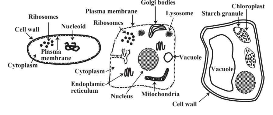
As noted in Section 7.2, there are two general classes of cells. Prokaryotic cells are those that make up bacteria and simple single-celled organisms that composed all of life on Earth for the first approximately 2 billion years of life on the planet. These cells are only about 1–2 micrometers in size, have only limited external appendages, and have relatively less (though still complex)internal structures. Eukaryotic cells compose all organisms other than bacteria, are typically 10μm or more in size, often have external appendages, and generally show well differentiated internal structures with numerous distinct parts. These cells appeared only about 1.5 billion years ago in the estimated 3.5 billion years that life has existed on Earth. Figure 12.1 represents prokaryotic cells and plant and animal eukaryotic cells.
Three features largely distinguish plant eukaryotic cells from animal cells in that the plant cells have a cell wall, a large central vacuole, and chloroplasts. The cell wall gives the plant cell strength and rigidity. The vacuole takes up most of the cell volume and allows contact with gases. The chloroplasts are sites in which chlorophyll uses light energy (hν) to synthesize carbohydrates as shown by the following reaction for the photosynthetic generation of glucose sugar:
\[\ce{6CO2 + 6H2O + (light energy, } h \nu \ce{) \rightarrow C6H12O6 (glucose) + 6O2}\]
Shown by the above reaction, photosynthesis was responsible for the greatest changes that the biosphere has ever caused in the atmosphere and geosphere. This occurred with the evolution of cyanobacteria (once thought to be algae and called “blue-green algae”) about 3 billion years ago, the first organisms capable of carrying out photosynthesis and producing oxygen, which for them was a waste product. This raised the oxygen content of the atmosphere from virtually zero to the current value of 21% (by volume of dry air). The result was conversion of the atmosphere to an oxidizing medium. Vast deposits of solid iron minerals now used for iron ore were formed when atmospheric oxygen reacted with dissolved Fe in the oceans to produce solid iron oxide.
\[\ce{4 Fe^{2+} + O2 + 4H2O \rightarrow 2Fe2O3 + 8 H^{+}}\]
Part of the oxygen generated by photosynthesis dissolved in water, where it was available for the development of organisms that used oxygen to metabolize organic matter. Whereas Earth’s surface had been a most inhospitable place for the existence of life, the oxygen released by photosynthesis enabled the formation of the ultraviolet-radiation-filtering layer of ozone (O3) in the stratosphere that made life possible outside the protective confines of water. Thus life became possible on Earth’s land surface, soil was formed, aided by the weathering action of organisms including cyanobacteria that grew on rock surfaces, plants growing in soil became well established, and animals developed. The huge changes made possible by the action of single-celled cyanobacteria carrying out photosynthesis are obvious.

Prokaryotic cells characteristic of bacteria are enclosed by strong cell walls composed largely of carbohydrates that hold the cells together. The plasma membrane controls passage of materials into and out of the cell and is the site of photosynthesis in photosynthetic bacteria. Gelatinous cytoplasm composed largely of protein and water fills the cell. There is not a defined nucleus, but the cell has a mass of genetic material (DNA) that composes a nucleoid. The DNA directs cell metabolism and reproduction. Proteins are made in the cell in ribosomes that are distributed around the cell interior. Ribosomes and other bodies in the prokaryotic cell are not enclosed by separate defined membranes as is the case with more complex eukaryotic cells.
Major Features of Eukaryotic Cells
Animal and plant cells shown in Figure 12.1 represent the two major kinds of eukaryotic cells that compose all organisms other than bacteria and cyanobacteria. The major features of eukaryotic cells include the following:
- Cell membrane, which encloses the cell and determines what enters and leaves the cell interior. The cell membrane has varying permeability for various substances so that one of its crucial functions is regulation of the passage of ions, nutrients, lipid-soluble (“fat-soluble”) substances, metabolic products, toxicants, and toxicant metabolites into and out of the cell interior thus protecting the contents of the cell from undesirable outside influences. One of the adverse effects of some toxicants is damage to the cell membrane causing the cell to function improperly.
- Cell nucleus, which controls cell function and the genetic material required for reproduction. Deoxyribonucleic acid (DNA) discussed in Section 7.6 is the key substance in the nucleus. Damage to DNA by foreign substances may cause mutations, cancer, birth defects, defective immune system function, and other toxiceffects.
- Cytoplasm composed of a water-soluble proteinaceous filler called cytosol fills the interior of the cell not occupied by the nucleus or other bodies. Bodies of cellular organelles, such as mitochondria or chloroplasts are suspended in the cytoplasm.
- Mitochondria mediate energy conversion and utilization where carbohydrates, proteins, and fats are broken down to yield carbon dioxide, water, and energy, which is then used by the cell. The best example of this is the oxidation of the sugar glucose, C6H12O6 in a process called cellular respiration (see Reaction 12.4.1).
- Ribosomes are involved in protein synthesis.
- Endoplasmic reticulum is the site of enzymatic metabolism of some toxicants.
- Lysosome, a type of organelle that contains potent substances capable of hydrolyzing and breaking down food material that enters the cell through a food vacuole.
- Golgi bodies are flattened bodies of material in some types of cells that serve to hold and release substances produced by the cells.
- Cell walls of provide stiffness and strength in cell walls composed primarily of cellulose (see Section 7.3 and Figure 7.3).
- Vacuoles inside plant cells often contain materials dissolved in water.
- Chloroplasts in plant cells that are the sites of photosynthesis, the chemical process which uses energy from sunlight to convert carbon dioxide and water to organic matter which is stored in the chloroplasts as starch grains.


