
14.3: Cancer
- Page ID
- 317982

\( \newcommand{\vecs}[1]{\overset { \scriptstyle \rightharpoonup} {\mathbf{#1}} } \)
\( \newcommand{\vecd}[1]{\overset{-\!-\!\rightharpoonup}{\vphantom{a}\smash {#1}}} \)
\( \newcommand{\id}{\mathrm{id}}\) \( \newcommand{\Span}{\mathrm{span}}\)
( \newcommand{\kernel}{\mathrm{null}\,}\) \( \newcommand{\range}{\mathrm{range}\,}\)
\( \newcommand{\RealPart}{\mathrm{Re}}\) \( \newcommand{\ImaginaryPart}{\mathrm{Im}}\)
\( \newcommand{\Argument}{\mathrm{Arg}}\) \( \newcommand{\norm}[1]{\| #1 \|}\)
\( \newcommand{\inner}[2]{\langle #1, #2 \rangle}\)
\( \newcommand{\Span}{\mathrm{span}}\)
\( \newcommand{\id}{\mathrm{id}}\)
\( \newcommand{\Span}{\mathrm{span}}\)
\( \newcommand{\kernel}{\mathrm{null}\,}\)
\( \newcommand{\range}{\mathrm{range}\,}\)
\( \newcommand{\RealPart}{\mathrm{Re}}\)
\( \newcommand{\ImaginaryPart}{\mathrm{Im}}\)
\( \newcommand{\Argument}{\mathrm{Arg}}\)
\( \newcommand{\norm}[1]{\| #1 \|}\)
\( \newcommand{\inner}[2]{\langle #1, #2 \rangle}\)
\( \newcommand{\Span}{\mathrm{span}}\) \( \newcommand{\AA}{\unicode[.8,0]{x212B}}\)
\( \newcommand{\vectorA}[1]{\vec{#1}} % arrow\)
\( \newcommand{\vectorAt}[1]{\vec{\text{#1}}} % arrow\)
\( \newcommand{\vectorB}[1]{\overset { \scriptstyle \rightharpoonup} {\mathbf{#1}} } \)
\( \newcommand{\vectorC}[1]{\textbf{#1}} \)
\( \newcommand{\vectorD}[1]{\overrightarrow{#1}} \)
\( \newcommand{\vectorDt}[1]{\overrightarrow{\text{#1}}} \)
\( \newcommand{\vectE}[1]{\overset{-\!-\!\rightharpoonup}{\vphantom{a}\smash{\mathbf {#1}}}} \)
\( \newcommand{\vecs}[1]{\overset { \scriptstyle \rightharpoonup} {\mathbf{#1}} } \)
\( \newcommand{\vecd}[1]{\overset{-\!-\!\rightharpoonup}{\vphantom{a}\smash {#1}}} \)
Cancer
Cancer has long been considered a cellular disease since cancers are composed of cells that grow without restraint in various areas of the body. Such growths of cancerous cells can replace normal cells or tissues causing severe malformations (such as with skin and bone cancers) and failure of internal organs which frequently leads to death. How do cells become cancerous? The development of cancer is an enormously complex process. For once a cell has started on the cancer path, it progresses through a series of steps, which continue long after the initial cause has disappeared.
Overview
There are about as many types of cancers as there are different types of cells in the body (over 100 types). Some cell types constantly divide and are replaced (such as skin and blood cells). Other types of cells rarely or never divide (such as bone cells and neurons). Sophisticated mechanisms exist in cells to control when, if, and how cells replicate. Cancer occurs when these mechanisms are lost and replication takes place in an uncontrolled and disorderly manner. It can arise when one cell or a small group of cells multiplies too many times because of damage to its DNA.
Recent research has begun to unravel the extremely complex pathogenesis of cancer. There is an intricate array of biochemical changes that take place within cells and between cells underlying the progression of cancer that transforms normal cells into cancerous cells. These biochemical changes lead a cell through a series of steps, changing it gradually from a normal to a cancer cell. The altered cell is no longer bound by the regulatory controls that govern the life and behavior of normal cells.
Cancer is not a single disease but a large group of diseases. The common aspect is that all cancers have the same basic property: they are composed of cells that no longer conform to the usual constraints on cell proliferation. In other words, they are uncontrolled growths of cells.
Terminology
The terminology associated with cancer can be confusing and may be used differently among the public and medical communities.
Here are definitions of the most frequently used cancer terms:
- Cancer — a malignant tumor that has the ability to metastasize or invade into surrounding tissues.
- Tumor — a general term for an uncontrolled growth of cells that becomes progressively worse with time. Tumors may be benign or malignant.
- Neoplasm — same as a tumor.
- Neoplasia — the growth of new tissue with abnormal and unregulated cellular proliferation.
- Benign Tumor — a tumor that does not metastasize or invade surrounding tissue.
- Malignant Tumor — a tumor that has the ability to metastasize or invade into surrounding tissues (same as cancer).
- Metastasis — ability to establish secondary tumor growth at a new location away from the original site.
- Carcinogenesis — the production of a carcinoma (epithelial cancer). Sometimes carcinogenesis is used as a general term for production of any type of tumor.
How are Cancers Named?
While most tumors are generally named in accordance with an internationally agreed-upon classification scheme, there are exceptions. Tumors are generally named and classified based on:
- The cell or tissue of origin
- Whether benign or malignant
Most tumor names end with the suffix "oma" which indicates a swelling or tissue enlargement. [Note: some terms ending with -oma are not cancers; for example, a hematoma is merely a swelling consisting of blood].
In naming tumors, qualifiers may be added in addition to the tissue of origin and structural features. For example, a "poorly-differentiated bronchogenic squamous cell carcinoma" is a malignant tumor (carcinoma) of squamous cell type (original cell type), which arose in the bronchi of the lung (site where the cancer started), and in which the cancer cells are poorly differentiated, meaning they have lost much of the normal appearance of squamous cells.
There are several historical exceptions to the standard nomenclature system, often based on their early and accepted use in the literature.
Examples include:
- Some tumors are named after the person that first described the tumor, for example, Wilms tumor (kidney tumor) and Hodgkin lymphoma (a specific form of lymphoid cancer).
- A few cancers are named for their physical characteristics such as pheochromocytomas (dark-colored tumors of the adrenal gland).
- A few cancers are composed of mixtures of cells, for example, fibrosarcoma and carcinosarcoma.
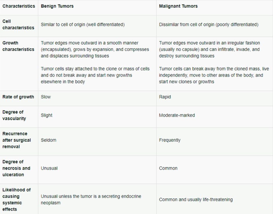
Most malignant tumors fall into one of two categories: carcinomas or sarcomas. The major differences between carcinomas and sarcomas are listed in Table \(\PageIndex{1}\):

Differences between Benign and Malignant Tumors
The biological and medical consequences of a tumor depend on whether it is benign or malignant.
Table \(\PageIndex{2}\) provides a comparison of the primary differences between benign and malignant tumors:
Common Sites for Cancer
Cancer can occur in almost any tissue or organ. Some cells and tissues are more likely to become cancerous than others are, particularly those cells that normally undergo proliferation to replace cells that have been lost due to injury or cell death. Those cells that don't proliferate (for example, neurons and heart muscle cells) rarely give rise to cancers. Figure \(\PageIndex{1}\) illustrates the most frequent occurrence of cancers in various body sites.
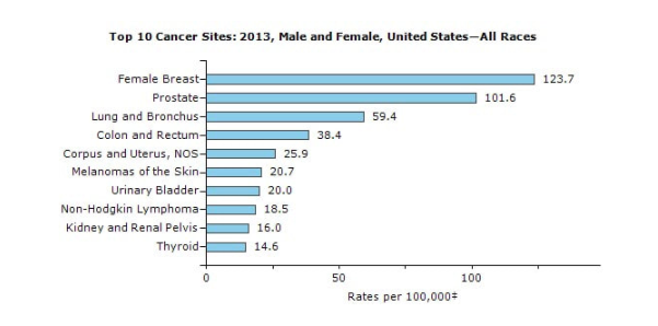
Figure \(\PageIndex{1}\). Top 10 Cancer Sites for Males and Females from All Races in the United States in 2013
(Image Source: CDC, https://nccd.cdc.gov/uscs/toptencancers.aspx)
‡ Rates are age-adjusted to the 2000 U.S. standard population (19 age groups – Census P25–1130)
While the prostate is the most common type of cancer that occurs in men, most survive with treatment. In contrast, other types of cancer are more often fatal. For example, the most common cancer, which causes death in men, even with treatment, is lung cancer. With women, a similar situation exists in that the breast is the most common site for cancer but more women die as a result of lung cancer.
What Do Cancers Look Like?
Cancer is a general term for more than 100 different cellular diseases, all with the same characteristic – the uncontrolled abnormal growth of cells in different parts of the body. Cancers appear in many forms. A few types are visible to the unaided eye but others grow inside the body and slowly destroy or replace internal tissues.
Skin Cancer
An example of a cancer that can be easily seen by the unaided eye is skin cancer. Skin cancers appear as raised, usually dark-colored, irregularly-shaped growths on the skin. As the cancer grows, it spreads to nearby areas of the skin. In advanced cases, the cancer metastasizes to lymph nodes and organs far away from the original site. The skin cancer illustrated in Figure \(\PageIndex{2}\) is known as a basal cell carcinoma. Melanomas and squamous cell carcinomas are other common skin cancers. Melanomas are usually the most malignant of the skin cancers.
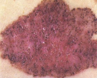
Figure \(\PageIndex{2}\). Photograph of basal cell carcinoma of the skin
(Image Source: NLM)
Other Cancers
Most cancers involve internal organs and require elaborate diagnostic tests to diagnose. Some large internal tumors can be felt or will push the skin outward and can be detected by noting abnormal bulges or an abnormal feel (for example, a hard area) to the body. Thyroid tumors, bone tumors, breast tumors, and testicular tumors are cancers that might be felt or observed by the patient. Other internal tumors may only be suspected based on diminished organ function (such as difficulty breathing with lung cancers), pain, bleeding (for example, blood in feces with colon cancer), weakness, or other unusual symptoms. To confirm the actual existence of a cancer may require diagnostic tests. This is especially the case where the cancer is not growing as a single large lump, but rather as a series of small tumors (metastatic foci) or when widely dispersed throughout the body (such as leukemia).
A few examples of internal cancers are presented in the following figures.
Liver Cancer
Numerous cancer nodules can be seen showing that much of the liver has been destroyed (Figure \(\PageIndex{3}\)).
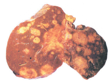
Figure \(\PageIndex{3}\). A liver with numerous cancer nodules
(Image Source: NLM)
Lung Cancer
An early developing squamous cell carcinoma can be seen growing in the middle of the lung (Figure \(\PageIndex{4}\)). As the cancer develops, it will consume more of the lung and metastasize to other areas of the body.
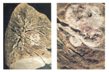
Figure \(\PageIndex{4}\). A cancerous lung
(Image Source: NLM)
Kidney Cancer
The photograph in Figure \(\PageIndex{5}\) shows the cancer has consumed much of the upper portion of the kidney.

Figure \(\PageIndex{5}\). A kidney with cancer
(Image Source: NLM)
Historical Changes in Incidence of Cancer
Cancer has been recognized in humans for centuries. However, the incidence of various types of cancer has changed since the mid-1900s. This is especially true for lung and stomach cancer. Deaths from lung cancer hit a peak in the early 1990s and have been slowly declining since 2001. During that same period, deaths from stomach cancer decreased substantially. Breast cancer caused more deaths than any other type cancer in women for many decades. However, when women began smoking cigarettes, deaths from lung cancer outpaced deaths from breast cancer. These changes in types and incidences of cancer reflect the increased longevity of people as well as personal habits and environmental changes.
Latency Period for Cancer Development
Cancer is a chronic condition, which develops gradually over a period of time, and may become a clinical concern many years following the initial exposure to a carcinogen. This period of time is referred to as the latency period. The latency period varies with the type of cancers and may be as short as a few years to over 30 years. For example, the latency period for leukemia after benzene or radiation exposure may only be five years. In contrast, the latency period may be 20–30 years for skin cancer after arsenic exposure and mesothelioma (cancer of the pleura around the lungs) after asbestos exposure.
Survival Time
Success in treating cancer varies greatly with the type of cancer with some cancers responding to treatment whereas others do not. For example, medical treatment of cancers of the pancreas, liver, esophagus, and lung are largely unsuccessful. In contrast, cancers of the thyroid, testes, and skin respond quite well to treatment. Table \(\PageIndex{3}\) shows the 5-year survival rate by cancer location.
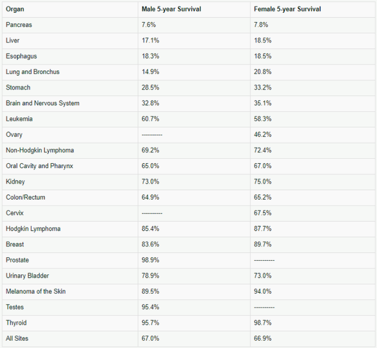
Table \(\PageIndex{3}\). Five year survival rate by primary cancer site (2006-2012)
(Source: Table 1. Surveillance, Epidemiology, and End Results Program (SEER), National Cancer Institute,
https://seer.cancer.gov/csr/1975_2013/results_merged/topic_survival.pdf)
What Causes Cancer?
A large number of industrial, pharmaceutical, and environmental chemicals have been identified as potential carcinogens by animal tests. Human epidemiology studies have confirmed that many are human carcinogens as well. However, while it is apparent that chemicals and radiation play a substantial role, it appears that lifestyle factors (such as diet, obesity, and smoking), and infections (such as hepatitis B, hepatitis C, and Human Papillomaviruses) are also major factors leading to the likelihood that a person will develop cancer. Additional factors that are involved in the development of cancer include aging and heredity.
Pathogenesis of Cancer
Carcinogenesis is a multi-step, multi-factorial genetic disease. All known tumors are composed of cells with genetic alterations that make them perform differently from their progenitor (parent) cells. The carcinogenesis process is very complex and unpredictable consisting of several phases and involving multiple genetic events (mutations) that take place over a very long period of time, at least 10 years for most types of cancer.
Cancer cells do not necessarily proliferate faster than their normal progenitors. In contrast to normal proliferating tissues where there is a strict and controlled balance between cell death and replacement, cancers grow and expand since more cancer cells are produced than die in a given time period. For a tumor to be detected it must attain a size of at least one cubic centimeter (about the size of a pea). This small tumor contains 100 million to a billion cells at that time. The development from a single cell to that size also means that the mass has doubled at least 30 times. During the long and active period of cell proliferation, the cancerous cells may have become aggressive in growth and have reverted to a less differentiated type cell that is not similar to the original cell type.
While knowledge of carcinogenesis continues to evolve, it is clear that there are at least three main phases in cancer development:
- Initiation
- Promotion/Conversion
- Progression
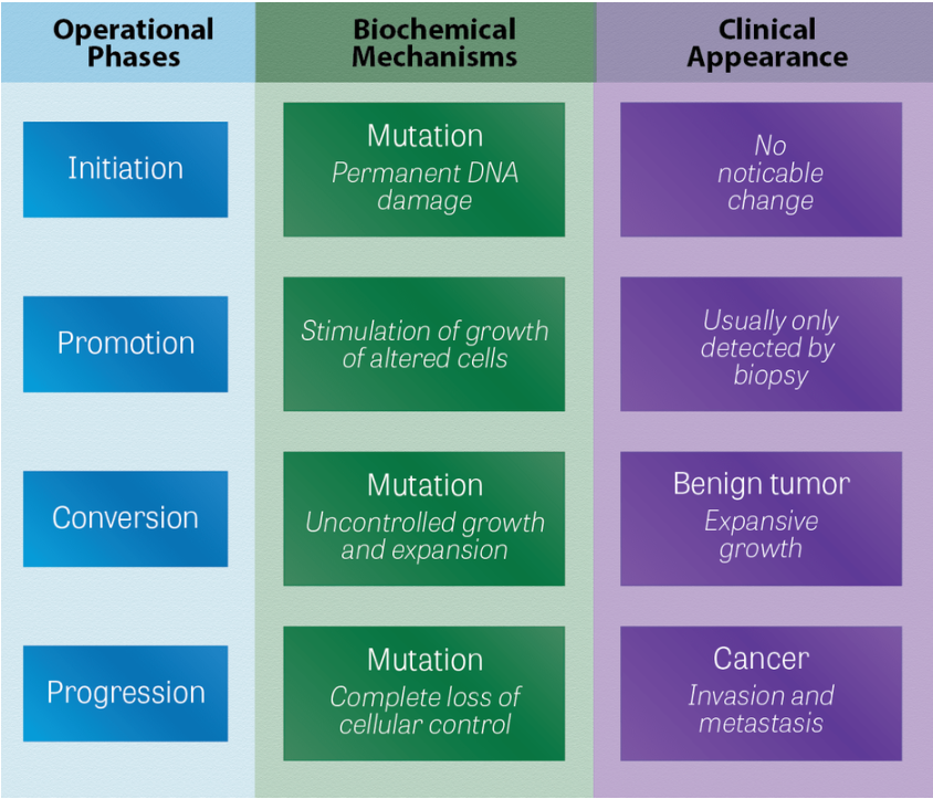
Figure \(\PageIndex{6}\). Phases of carcinogenesis
(Image Source: NLM)
1. Initiation
The initiation phase consists of the alteration of the DNA (mutation) of a normal cell, which is an irreversible change. The initiated cell has developed a capacity for individual growth. At this time, the initiated cell is indistinguishable from other similar cells in the tissue. The initiating event can consist of a single exposure to a carcinogenic agent or, in some cases, it may be an inherited genetic defect.
- An example is retinoblastoma in which some children who develop the disease may have inherited an altered copy of the gene involved and are at risk of passing the altered gene to successive generations.
The initiated cell, whether inherited or newly mutated, may remain dormant for months to years and, unless a promoting event occurs, may never develop into a clinical cancer case.
2. Promotion/Conversion
The promotion/conversion phase is the second major step in the carcinogenesis process in which specific agents (referred to as promoters) enhance the further development of the initiated cells. Promoters often, but not always, interact with the cell's DNA and influence the further expression of the mutated DNA so that the initiated cell proliferates and progresses further through the carcinogenesis process. The clone of proliferating cells in this stage takes a form consistent with a benign tumor. The mass of cells remains as a cohesive group and physically keeps in contact with each other.
3. Progression
Progression is the third recognized step and is associated with the development of the initiated cell into a biologically malignant cell population. In this stage, a portion of the benign tumor cells may be converted into malignant forms so that a true cancer has evolved. Individual cells in this final stage can break away and start new clones of growth distant from the original site of development of the tumor. This is known as metastasis.
Genetic Activity
While the three-stage pathogenesis scheme describes the basic sequence of events in the carcinogenesis process, the actual events that take place in these various steps are due to activities of specific genes within the DNA of the cells. Cellular DNA contains two types of genes:
- Structural genes direct the production of specific proteins within the cell.
- Regulatory genes control the activity of the structural genes and direct the proliferation process of the cell.
The three classes of regulatory genes considered to have major roles in the carcinogenesis process are known as:
- Proto-oncogenes
- Oncogenes
- Suppressor genes
Proto-oncogenes are normal cellular genes that encode and instruct the production of the regulatory proteins and growth factors within the cell or its membrane. The proteins encoded by proto-oncogenes are necessary for normal cellular cell growth and differentiation. Activation of a proto-oncogene can cause the alteration in the normal growth and differentiation of cells, which leads to neoplasia. Several agents can activate proto-oncogenes. This is the result of point mutations or by DNA rearrangements of the proto-oncogenes. The product of this proto-oncogene activation is an oncogene. Many proto-oncogenes have been identified and have usually been named after the source of their discovery, for example, the KRAS proto-oncogene was named for the discovery using the Kirsten rat sarcoma virus. HRAS, MYC, MYB, and SRC are other examples of proto-oncogenes. The proto-oncogenes are not specific for the original species but have been found in many other species, including humans. These proto-oncogenes are present in many cells but remain dormant until activated. Either a point mutation or chromosomal damage of various types can induce activation. Once activated they become an oncogene.
Oncogenes are altered or misdirected proto-oncogenes which now have the ability to direct the production of proteins within the cell that can change or transform the normal cell into a neoplastic cell. Most oncogenes differ from their proto-oncogenes by a single point mutation located at a specific codon (a group of three DNA bases that encodes for a specific amino acid) of a chromosome. The altered DNA in the oncogene results in the production of an abnormal protein that can alter cell growth and differentiation. It appears that a single activated oncogene is not sufficient for the growth and progression of a cell and its offspring to form a cancerous growth. However, it is a major step in the carcinogenesis process.
Tumor suppressor genes, sometimes referred to as anti-oncogenes, are present in normal cells and serve to counteract and change the proto-oncogenes and altered proteins that they are responsible for. The tumor suppressor genes serve to prevent a cell with damaged DNA from proliferating and evolving into an uncontrolled growth. They actively function to effectively oppose the action of an oncogene. If a tumor suppressor gene is inactivated (usually by a point mutation), its control over the oncogene and transformed cell may be lost. Thus the tumor-potential cell can now grow without restraint and is free of the normal cellular regulatory control. The suppressor gene most frequently altered in human tumors is the p53 gene. Damaged p53 genes have been identified in over 50% of human cancers.
The p53 gene normally halts cell division and stimulates repair enzymes to rebuild and restore the damaged regions of the DNA. If the damage is too extensive, the p53 commands the cell to self-destruct. An altered p53 is incapable of these defensive actions and cannot prevent the cell with damaged DNA from dividing and proliferating in an erratic and uncontrolled manner. This is the essence of cancer.
This section represents only a brief overview of an enormously complex process for which knowledge is continuously evolving with the tools of molecular biology. New factors are continuously being identified; however, many pieces of this cancer puzzle remain elusive at this time.
1) A body growth with the ability to metastasize or invade into surrounding tissues is known as a:
a) Benign tumor
b) Malignant tumor
c) Hyperplasia
- Answer
-
Malignant tumor - This is the correct answer.
A malignant tumor that has the ability to metastasize or invade into surrounding tissues. It is the same as cancer.
2) Most cancers are thought to be due to the following:
a) Infections
b) Food additives
c) Lifestyle factors
d) Pollution
- Answer
-
Lifestyle factors - This is the correct answer.
Lifestyle (including diet, tobacco use, reproductive and sexual behavior, and alcohol consumption) is considered to cause about 75% of all cancers.
3) The initial stage in carcinogenesis in which there is an alteration of the DNA (mutation) is referred to as the:
a) Progression stage
b) Promotion stage
c) Initiation stage
- Answer
-
Initiation stage - This is the correct answer.
The initiation phase consists of the alteration of the DNA (mutation) of a normal cell, which is irreversible change.
4) The cellular gene which is present in most normal cells and serves as a balance to the genes for tumor expression is known as a:
a) Tumor suppressor gene
b) Oncogene
c) Proto-oncogene
- Answer
-
Tumor suppressor gene - This is the correct answer.
Tumor suppressor genes, sometimes referred to as anti-oncogenes, are present in normal cells and serve as a balance to the genes for expression or proto-oncogenes.