3.4: Different Cytotoxicity Assays
- Page ID
- 308338
\( \newcommand{\vecs}[1]{\overset { \scriptstyle \rightharpoonup} {\mathbf{#1}} } \)
\( \newcommand{\vecd}[1]{\overset{-\!-\!\rightharpoonup}{\vphantom{a}\smash {#1}}} \)
\( \newcommand{\dsum}{\displaystyle\sum\limits} \)
\( \newcommand{\dint}{\displaystyle\int\limits} \)
\( \newcommand{\dlim}{\displaystyle\lim\limits} \)
\( \newcommand{\id}{\mathrm{id}}\) \( \newcommand{\Span}{\mathrm{span}}\)
( \newcommand{\kernel}{\mathrm{null}\,}\) \( \newcommand{\range}{\mathrm{range}\,}\)
\( \newcommand{\RealPart}{\mathrm{Re}}\) \( \newcommand{\ImaginaryPart}{\mathrm{Im}}\)
\( \newcommand{\Argument}{\mathrm{Arg}}\) \( \newcommand{\norm}[1]{\| #1 \|}\)
\( \newcommand{\inner}[2]{\langle #1, #2 \rangle}\)
\( \newcommand{\Span}{\mathrm{span}}\)
\( \newcommand{\id}{\mathrm{id}}\)
\( \newcommand{\Span}{\mathrm{span}}\)
\( \newcommand{\kernel}{\mathrm{null}\,}\)
\( \newcommand{\range}{\mathrm{range}\,}\)
\( \newcommand{\RealPart}{\mathrm{Re}}\)
\( \newcommand{\ImaginaryPart}{\mathrm{Im}}\)
\( \newcommand{\Argument}{\mathrm{Arg}}\)
\( \newcommand{\norm}[1]{\| #1 \|}\)
\( \newcommand{\inner}[2]{\langle #1, #2 \rangle}\)
\( \newcommand{\Span}{\mathrm{span}}\) \( \newcommand{\AA}{\unicode[.8,0]{x212B}}\)
\( \newcommand{\vectorA}[1]{\vec{#1}} % arrow\)
\( \newcommand{\vectorAt}[1]{\vec{\text{#1}}} % arrow\)
\( \newcommand{\vectorB}[1]{\overset { \scriptstyle \rightharpoonup} {\mathbf{#1}} } \)
\( \newcommand{\vectorC}[1]{\textbf{#1}} \)
\( \newcommand{\vectorD}[1]{\overrightarrow{#1}} \)
\( \newcommand{\vectorDt}[1]{\overrightarrow{\text{#1}}} \)
\( \newcommand{\vectE}[1]{\overset{-\!-\!\rightharpoonup}{\vphantom{a}\smash{\mathbf {#1}}}} \)
\( \newcommand{\vecs}[1]{\overset { \scriptstyle \rightharpoonup} {\mathbf{#1}} } \)
\(\newcommand{\longvect}{\overrightarrow}\)
\( \newcommand{\vecd}[1]{\overset{-\!-\!\rightharpoonup}{\vphantom{a}\smash {#1}}} \)
\(\newcommand{\avec}{\mathbf a}\) \(\newcommand{\bvec}{\mathbf b}\) \(\newcommand{\cvec}{\mathbf c}\) \(\newcommand{\dvec}{\mathbf d}\) \(\newcommand{\dtil}{\widetilde{\mathbf d}}\) \(\newcommand{\evec}{\mathbf e}\) \(\newcommand{\fvec}{\mathbf f}\) \(\newcommand{\nvec}{\mathbf n}\) \(\newcommand{\pvec}{\mathbf p}\) \(\newcommand{\qvec}{\mathbf q}\) \(\newcommand{\svec}{\mathbf s}\) \(\newcommand{\tvec}{\mathbf t}\) \(\newcommand{\uvec}{\mathbf u}\) \(\newcommand{\vvec}{\mathbf v}\) \(\newcommand{\wvec}{\mathbf w}\) \(\newcommand{\xvec}{\mathbf x}\) \(\newcommand{\yvec}{\mathbf y}\) \(\newcommand{\zvec}{\mathbf z}\) \(\newcommand{\rvec}{\mathbf r}\) \(\newcommand{\mvec}{\mathbf m}\) \(\newcommand{\zerovec}{\mathbf 0}\) \(\newcommand{\onevec}{\mathbf 1}\) \(\newcommand{\real}{\mathbb R}\) \(\newcommand{\twovec}[2]{\left[\begin{array}{r}#1 \\ #2 \end{array}\right]}\) \(\newcommand{\ctwovec}[2]{\left[\begin{array}{c}#1 \\ #2 \end{array}\right]}\) \(\newcommand{\threevec}[3]{\left[\begin{array}{r}#1 \\ #2 \\ #3 \end{array}\right]}\) \(\newcommand{\cthreevec}[3]{\left[\begin{array}{c}#1 \\ #2 \\ #3 \end{array}\right]}\) \(\newcommand{\fourvec}[4]{\left[\begin{array}{r}#1 \\ #2 \\ #3 \\ #4 \end{array}\right]}\) \(\newcommand{\cfourvec}[4]{\left[\begin{array}{c}#1 \\ #2 \\ #3 \\ #4 \end{array}\right]}\) \(\newcommand{\fivevec}[5]{\left[\begin{array}{r}#1 \\ #2 \\ #3 \\ #4 \\ #5 \\ \end{array}\right]}\) \(\newcommand{\cfivevec}[5]{\left[\begin{array}{c}#1 \\ #2 \\ #3 \\ #4 \\ #5 \\ \end{array}\right]}\) \(\newcommand{\mattwo}[4]{\left[\begin{array}{rr}#1 \amp #2 \\ #3 \amp #4 \\ \end{array}\right]}\) \(\newcommand{\laspan}[1]{\text{Span}\{#1\}}\) \(\newcommand{\bcal}{\cal B}\) \(\newcommand{\ccal}{\cal C}\) \(\newcommand{\scal}{\cal S}\) \(\newcommand{\wcal}{\cal W}\) \(\newcommand{\ecal}{\cal E}\) \(\newcommand{\coords}[2]{\left\{#1\right\}_{#2}}\) \(\newcommand{\gray}[1]{\color{gray}{#1}}\) \(\newcommand{\lgray}[1]{\color{lightgray}{#1}}\) \(\newcommand{\rank}{\operatorname{rank}}\) \(\newcommand{\row}{\text{Row}}\) \(\newcommand{\col}{\text{Col}}\) \(\renewcommand{\row}{\text{Row}}\) \(\newcommand{\nul}{\text{Nul}}\) \(\newcommand{\var}{\text{Var}}\) \(\newcommand{\corr}{\text{corr}}\) \(\newcommand{\len}[1]{\left|#1\right|}\) \(\newcommand{\bbar}{\overline{\bvec}}\) \(\newcommand{\bhat}{\widehat{\bvec}}\) \(\newcommand{\bperp}{\bvec^\perp}\) \(\newcommand{\xhat}{\widehat{\xvec}}\) \(\newcommand{\vhat}{\widehat{\vvec}}\) \(\newcommand{\uhat}{\widehat{\uvec}}\) \(\newcommand{\what}{\widehat{\wvec}}\) \(\newcommand{\Sighat}{\widehat{\Sigma}}\) \(\newcommand{\lt}{<}\) \(\newcommand{\gt}{>}\) \(\newcommand{\amp}{&}\) \(\definecolor{fillinmathshade}{gray}{0.9}\)- 1: Know different types of cytotoxicity assays.
- 2: Know how different cytotoxicity assays are used when cells are exposed to toxicants or mutagens.
The goal of cytotoxicity assay is to determine whether any chemicals or drugs will do any toxic effect or load on milieu or genetic material of the cells caused for lethality of the cells or caused for different diseases.
The following are the different types of cytotoxicity assays:
- DNA fragmentation/ladder assay
- Comet assay
- Necrosis assay
- Enzyme assay
- Proteomics assay
- Expression array assay
4.1: DNA Fragmentation/Ladder Assay
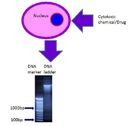
DNA fragmentation or ladder assay are used to know the fragmented DNA of the cells caused by chemicals or drugs. Fragmented DNA can be separated by agarose gel electrophoresis and can be visualized as “ladder” by ethidium bromide staining. The evaluation of cytotoxicity through cell death is an acceptable common assessment.
Ladder assays are performed for the following reasons:
- to simply characterize the toxicity of the chemicals or drugs in cells, or
- to determine the maximum doses of the test chemicals or drugs that can be used for cells without causing too much cell death.

4.2: Comet Assay
The Comet Assay (single cell gel electrophoresis /SCGE) is used to detect DNA damage by using a micro gel electrophoresis. The image of the damaged DNA shows a comet with head and tail. The analysis of image for comet assay is calculated for the “tail length” of the comet which is the measurement from the point of highest intensity within the comet head as well as the “tail moment” which is the product of the tail length and the fraction of total DNA present within the tail.

4.3: Necrosis Assay
The necrosis assay is performed by flow cytometry analysis with staining of Annexin V and propidium iodide (PI) in the cells. The cells are considered in the stage of necrosis if the cells lose membrane integrity and die promptly due to cell lysis when exposed to chemicals, drugs, toxins or foreign antigens. In necrosis, cells show swelling, loss of membrane integrity and disruption of metabolism. Cells with necrosis do not go to stage of apoptosis, apoptotic cell may undergo secondary necrosis. These necrotic cells will shut down metabolism, lose membrane integrity, lyse and formed cell injury autolysis.
The flow cytometric analysis for necrosis assay showed that the cells stained positive for both FITC Annexin V and PI are in the end stage of apoptosis and are undergoing to the stage of necrosis as dead cells stained PI positive. Cells that stain negative for both FITC Annexin V and PI are alive and not undergoing apoptosis or necrosis.

4.4: Enzyme Assay
The enzyme assay is used to monitor passaging of lactate dehydrogenase (LDH), due to loss of cell membrane integrity when cells are exposed to cytotoxic compounds. LDH reduces NAD to NADH which generates a color change by interaction with a specific probe (Figure 4a). In other enzyme assay, Adenosine triphosphate (ATP )-based assay combined with bioluminescent assay are used to measure cytotoxicity of the cells in which ATP is the reagent for the luciferase reaction.

4.5: Proteomics Assay
The proteomic assay is performed to know the mechanism of cellular toxicity by measuring expression of a specific protein which may consider as a biomarker for particular toxic mechanism or cellular toxicity signaling pathway. Immunofluorescence, immunoprecipitation and immunoblot assay are mainly used to know the effect of toxicants in cellular toxicity signaling pathway or mechanism.

4.6: Expression Array Assay
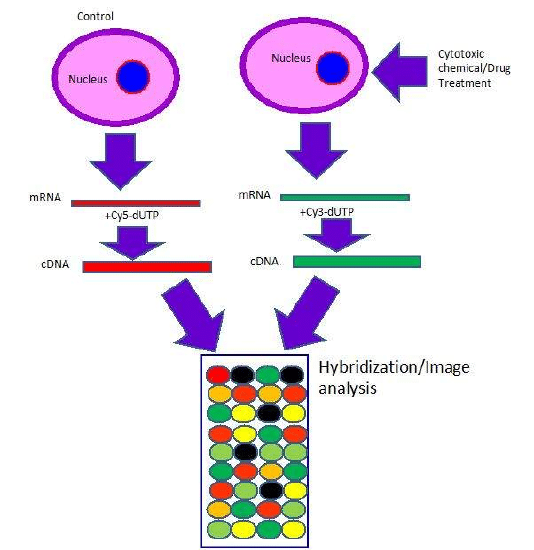
The expression array is the chip based microarray of more gene expressions (finger print of genes) by the effect of cellular toxicants. This is a rapid and sensitive detection method which allows to detect all toxicological end points at wide range of molecular level changes in the cell at single assay. The microarray process can be divided into two main parts. First is the printing of known gene sequences onto glass slides or other solid support followed by hybridization of fluorescently labeled cDNA (containing the unknown sequences to be interrogated) to the known genes immobilized on the glass slide. After hybridization, arrays are scanned using a fluorescent microarray scanner. Analyzing the relative fluorescent intensity of different genes provides a measure of the differences in gene expression.

Topic 4: Key Points
In this section, we explored the following main points:
- 1: Different types of cytotoxicity assays.
- 2: How different cytotoxicity assay namely DNA fragmentation/ladder assay, Comet assay, Necrosis assay, Enzyme assay, Proteomics assay, and Expression array assay are used when cells are exposed to cytotoxic agents.
1. The cells are considered in the stage of necrosis, if the cells lose membrane integrity and die promptly due to cell lysis when exposed to chemicals, drugs, toxins or foreign antigens.
True
False
- Answer
-
True
2. The Comet Assay is used to detect DNA damage by using a micro gel electrophoresis:
True
False
- Answer
-
True
3. The expression array is the chip based microarray of more gene expressions (finger print of genes) by the effect of cellular toxicants.
True
False
- Answer
-
True
4. Ladder assay are performed for the following reasons: 1) to simply characterize the toxicity of the chemicals or drugs in cells, or 2) to determine the maximum doses of the test chemicals or drugs that can be used for cells without causing too much cell death.
True
False
- Answer
-
True
5. What are the proteomic assays to know the effect of toxicants in cellular toxicity signaling pathway or mechanism?
Immunofluorescence assay
ring
Immunoprecipitation assay
Immunoblot assay
All of the above
- Answer
-
All of the above


