
1.4: Cellular Response to Toxicant-Induced Injury
- Page ID
- 308321

\( \newcommand{\vecs}[1]{\overset { \scriptstyle \rightharpoonup} {\mathbf{#1}} } \)
\( \newcommand{\vecd}[1]{\overset{-\!-\!\rightharpoonup}{\vphantom{a}\smash {#1}}} \)
\( \newcommand{\dsum}{\displaystyle\sum\limits} \)
\( \newcommand{\dint}{\displaystyle\int\limits} \)
\( \newcommand{\dlim}{\displaystyle\lim\limits} \)
\( \newcommand{\id}{\mathrm{id}}\) \( \newcommand{\Span}{\mathrm{span}}\)
( \newcommand{\kernel}{\mathrm{null}\,}\) \( \newcommand{\range}{\mathrm{range}\,}\)
\( \newcommand{\RealPart}{\mathrm{Re}}\) \( \newcommand{\ImaginaryPart}{\mathrm{Im}}\)
\( \newcommand{\Argument}{\mathrm{Arg}}\) \( \newcommand{\norm}[1]{\| #1 \|}\)
\( \newcommand{\inner}[2]{\langle #1, #2 \rangle}\)
\( \newcommand{\Span}{\mathrm{span}}\)
\( \newcommand{\id}{\mathrm{id}}\)
\( \newcommand{\Span}{\mathrm{span}}\)
\( \newcommand{\kernel}{\mathrm{null}\,}\)
\( \newcommand{\range}{\mathrm{range}\,}\)
\( \newcommand{\RealPart}{\mathrm{Re}}\)
\( \newcommand{\ImaginaryPart}{\mathrm{Im}}\)
\( \newcommand{\Argument}{\mathrm{Arg}}\)
\( \newcommand{\norm}[1]{\| #1 \|}\)
\( \newcommand{\inner}[2]{\langle #1, #2 \rangle}\)
\( \newcommand{\Span}{\mathrm{span}}\) \( \newcommand{\AA}{\unicode[.8,0]{x212B}}\)
\( \newcommand{\vectorA}[1]{\vec{#1}} % arrow\)
\( \newcommand{\vectorAt}[1]{\vec{\text{#1}}} % arrow\)
\( \newcommand{\vectorB}[1]{\overset { \scriptstyle \rightharpoonup} {\mathbf{#1}} } \)
\( \newcommand{\vectorC}[1]{\textbf{#1}} \)
\( \newcommand{\vectorD}[1]{\overrightarrow{#1}} \)
\( \newcommand{\vectorDt}[1]{\overrightarrow{\text{#1}}} \)
\( \newcommand{\vectE}[1]{\overset{-\!-\!\rightharpoonup}{\vphantom{a}\smash{\mathbf {#1}}}} \)
\( \newcommand{\vecs}[1]{\overset { \scriptstyle \rightharpoonup} {\mathbf{#1}} } \)
\(\newcommand{\longvect}{\overrightarrow}\)
\( \newcommand{\vecd}[1]{\overset{-\!-\!\rightharpoonup}{\vphantom{a}\smash {#1}}} \)
\(\newcommand{\avec}{\mathbf a}\) \(\newcommand{\bvec}{\mathbf b}\) \(\newcommand{\cvec}{\mathbf c}\) \(\newcommand{\dvec}{\mathbf d}\) \(\newcommand{\dtil}{\widetilde{\mathbf d}}\) \(\newcommand{\evec}{\mathbf e}\) \(\newcommand{\fvec}{\mathbf f}\) \(\newcommand{\nvec}{\mathbf n}\) \(\newcommand{\pvec}{\mathbf p}\) \(\newcommand{\qvec}{\mathbf q}\) \(\newcommand{\svec}{\mathbf s}\) \(\newcommand{\tvec}{\mathbf t}\) \(\newcommand{\uvec}{\mathbf u}\) \(\newcommand{\vvec}{\mathbf v}\) \(\newcommand{\wvec}{\mathbf w}\) \(\newcommand{\xvec}{\mathbf x}\) \(\newcommand{\yvec}{\mathbf y}\) \(\newcommand{\zvec}{\mathbf z}\) \(\newcommand{\rvec}{\mathbf r}\) \(\newcommand{\mvec}{\mathbf m}\) \(\newcommand{\zerovec}{\mathbf 0}\) \(\newcommand{\onevec}{\mathbf 1}\) \(\newcommand{\real}{\mathbb R}\) \(\newcommand{\twovec}[2]{\left[\begin{array}{r}#1 \\ #2 \end{array}\right]}\) \(\newcommand{\ctwovec}[2]{\left[\begin{array}{c}#1 \\ #2 \end{array}\right]}\) \(\newcommand{\threevec}[3]{\left[\begin{array}{r}#1 \\ #2 \\ #3 \end{array}\right]}\) \(\newcommand{\cthreevec}[3]{\left[\begin{array}{c}#1 \\ #2 \\ #3 \end{array}\right]}\) \(\newcommand{\fourvec}[4]{\left[\begin{array}{r}#1 \\ #2 \\ #3 \\ #4 \end{array}\right]}\) \(\newcommand{\cfourvec}[4]{\left[\begin{array}{c}#1 \\ #2 \\ #3 \\ #4 \end{array}\right]}\) \(\newcommand{\fivevec}[5]{\left[\begin{array}{r}#1 \\ #2 \\ #3 \\ #4 \\ #5 \\ \end{array}\right]}\) \(\newcommand{\cfivevec}[5]{\left[\begin{array}{c}#1 \\ #2 \\ #3 \\ #4 \\ #5 \\ \end{array}\right]}\) \(\newcommand{\mattwo}[4]{\left[\begin{array}{rr}#1 \amp #2 \\ #3 \amp #4 \\ \end{array}\right]}\) \(\newcommand{\laspan}[1]{\text{Span}\{#1\}}\) \(\newcommand{\bcal}{\cal B}\) \(\newcommand{\ccal}{\cal C}\) \(\newcommand{\scal}{\cal S}\) \(\newcommand{\wcal}{\cal W}\) \(\newcommand{\ecal}{\cal E}\) \(\newcommand{\coords}[2]{\left\{#1\right\}_{#2}}\) \(\newcommand{\gray}[1]{\color{gray}{#1}}\) \(\newcommand{\lgray}[1]{\color{lightgray}{#1}}\) \(\newcommand{\rank}{\operatorname{rank}}\) \(\newcommand{\row}{\text{Row}}\) \(\newcommand{\col}{\text{Col}}\) \(\renewcommand{\row}{\text{Row}}\) \(\newcommand{\nul}{\text{Nul}}\) \(\newcommand{\var}{\text{Var}}\) \(\newcommand{\corr}{\text{corr}}\) \(\newcommand{\len}[1]{\left|#1\right|}\) \(\newcommand{\bbar}{\overline{\bvec}}\) \(\newcommand{\bhat}{\widehat{\bvec}}\) \(\newcommand{\bperp}{\bvec^\perp}\) \(\newcommand{\xhat}{\widehat{\xvec}}\) \(\newcommand{\vhat}{\widehat{\vvec}}\) \(\newcommand{\uhat}{\widehat{\uvec}}\) \(\newcommand{\what}{\widehat{\wvec}}\) \(\newcommand{\Sighat}{\widehat{\Sigma}}\) \(\newcommand{\lt}{<}\) \(\newcommand{\gt}{>}\) \(\newcommand{\amp}{&}\) \(\definecolor{fillinmathshade}{gray}{0.9}\)After completing this lesson, you will be able to:
- 1: Describe the range of cellular injury that may occur following exposure to a toxicant.
- 2: Discuss the major mechanisms of toxicant-induced cell death.
- 3: Compare the processes of necrosis and apoptosis in terms of inciting causes and the cellular changes that occur with each.
 Toxicants can exert a variety of effects at the molecular level that have significant repercussions at the cellular, tissue and organ levels. The effects of a toxic exposure can range from reversible cellular dysfunction to irreversible cellular injury to cell death, all of which can alter normal organ function and have significant impact on the health and well-being of the body as a whole. The ability of cells, tissues and organs to overcome the effect of a toxicant through repair and/or adaptation will dictate the ultimate outcome of a toxic exposure.


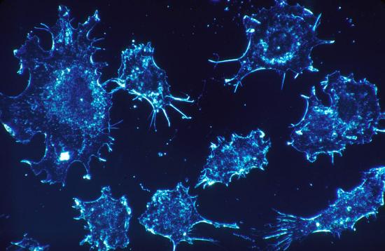
Following toxic insult, cells have a limited repertoire of responses. Nonlethal cell injury may lead to cellular degeneration, seen microscopically as swelling of the cells. Toxicant-injured cells may accumulate water, lipid, pigments, glycogen or metabolic waste products due to impairment of normal maintenance functions. Accumulation of lipids in hepatocytes, termed steatosis or hepatic lipidosis, is a common toxic effect seen in cases of alcohol-related liver disease. Degenerative changes in cells are often reversible if the inciting cause is removed. When cell injury proceeds beyond the self-repair capability of the cell, cell death ensues.
Mechanisms of Cell Death
Major mechanisms of toxicant-induced cell death include disruption of cell membrane structure and/or function, loss of cellular maintenance functions, and impairment of cellular energy production. Loss of membrane structural or functional integrity can result in uncontrolled passage of water, ions and other compounds into or out of the cell. The subsequent loss of normal cytosolic environment interferes with normal biochemical processes necessary for cell function and/or survival. Loss of ability to synthesize proteins and other macromolecules impedes maintenance of organelles and enzymatic pathways vital to cellular survival. Impairment of cellular energy production generally occurs when toxic effects alter mitochondrial function and/or structure and can lead to cell death due to failure to produce sufficient ATP to power essential cellular functions.
Necrosis
Necrosis is the term used to describe cell death due to irreversible injury. Necrotic cells undergo degenerative processes including swelling of organelles, loss of organelle function, oxidative and hydrolytic degradation of intracellular membranes and macromolecules by electophiles and free radicals, and, ultimately, lysis (loss of cellular constituents to surrounding tissues due to cell membrane rupture). Necrosis generally results in the generation of an inflammatory response as cellular components and free radicals that are released to the extracellular matrix attract inflammatory cells.
Apoptosis
In contrast, apoptosis (sometimes nicknamed “cell suicide” or “programmed cell death”) is a more orderly form of cell death. Apoptosis is an active process involving activation of specific enzymes which triggers the systematic fragmentation of cell constituents into blebs of cell membrane that pinch off of the main cell to form apoptotic bodies. During this fragmentation, the cell continues to produce energy and proteins, unlike necrosis where organelle and energy production cease prior to cellular fragmentation. The end result of apoptosis is numerous apoptotic bodies, each composed of a cellular membrane surrounding intact and functional cellular components. Apoptosis can be triggered by various forms of oxidative stress, particularly the presence of excessive oxygen-derived free radicals, due to excessive free radical generation and/or to lack or exhaustion of endogenous antioxidants. Because intracellular components are not spilled into the extracellular matrix, apoptosis generally does not incite an inflammatory response; instead the apoptotic bodies are removed by local phagocytes.

Topic 4: Key Points
In this section, we explored the following main points:
- 1: Toxicant-induced cellular injury can range from reversible cellular dysfunction to irreversible cellular injury to cell death.
- 2: Cellular responses to toxic injury may include cellular degeneration and accumulation of substrates within the cell.
- Steatosis is lipid accumulation within hepatocytes and is a common toxic effect secondary to alcohol exposure.
- 3: Major mechanisms of cell death include disruption of cell membrane structure and/or function, loss of cellular maintenance functions, and impairment of cellular energy production.
- 4: Necrosis is cell death resulting from cessation of organelle function, degradation of intracellular structures and culminating in lysis of the cell and attraction of inflammatory cells.
- 5: Apoptosis is an active form of cell death whereby cellular function is maintained as the cell components are compartmentalized and packaged into apoptotic bodies that pinch off of the main cell.
- Apoptosis can be triggered by oxidative stresses caused by oxygen-derived free radicals.
- Apoptosis is not generally associated with an inflammatory response.
1. The accumulation of excess lipid within liver cells:
- Answer
-
Steatosis
2. Cellular degeneration secondary to a toxic insult is seen microscopically as:
- Answer
-
Cellular Swelling
3. Loss of organelle function, hydrolytic degradation of intracellular membranes and lysis of cells are characteristics of_______.
- Answer
-
necrosis
4. The orderly decommissioning of cellular organelles without loss of energy and protein synthesis leading to fragmentation into membrane-bound packets is characteristic of_____.
- Answer
-
apoptosis