
C. Working Electrodes
- Page ID
- 61543

\( \newcommand{\vecs}[1]{\overset { \scriptstyle \rightharpoonup} {\mathbf{#1}} } \)
\( \newcommand{\vecd}[1]{\overset{-\!-\!\rightharpoonup}{\vphantom{a}\smash {#1}}} \)
\( \newcommand{\id}{\mathrm{id}}\) \( \newcommand{\Span}{\mathrm{span}}\)
( \newcommand{\kernel}{\mathrm{null}\,}\) \( \newcommand{\range}{\mathrm{range}\,}\)
\( \newcommand{\RealPart}{\mathrm{Re}}\) \( \newcommand{\ImaginaryPart}{\mathrm{Im}}\)
\( \newcommand{\Argument}{\mathrm{Arg}}\) \( \newcommand{\norm}[1]{\| #1 \|}\)
\( \newcommand{\inner}[2]{\langle #1, #2 \rangle}\)
\( \newcommand{\Span}{\mathrm{span}}\)
\( \newcommand{\id}{\mathrm{id}}\)
\( \newcommand{\Span}{\mathrm{span}}\)
\( \newcommand{\kernel}{\mathrm{null}\,}\)
\( \newcommand{\range}{\mathrm{range}\,}\)
\( \newcommand{\RealPart}{\mathrm{Re}}\)
\( \newcommand{\ImaginaryPart}{\mathrm{Im}}\)
\( \newcommand{\Argument}{\mathrm{Arg}}\)
\( \newcommand{\norm}[1]{\| #1 \|}\)
\( \newcommand{\inner}[2]{\langle #1, #2 \rangle}\)
\( \newcommand{\Span}{\mathrm{span}}\) \( \newcommand{\AA}{\unicode[.8,0]{x212B}}\)
\( \newcommand{\vectorA}[1]{\vec{#1}} % arrow\)
\( \newcommand{\vectorAt}[1]{\vec{\text{#1}}} % arrow\)
\( \newcommand{\vectorB}[1]{\overset { \scriptstyle \rightharpoonup} {\mathbf{#1}} } \)
\( \newcommand{\vectorC}[1]{\textbf{#1}} \)
\( \newcommand{\vectorD}[1]{\overrightarrow{#1}} \)
\( \newcommand{\vectorDt}[1]{\overrightarrow{\text{#1}}} \)
\( \newcommand{\vectE}[1]{\overset{-\!-\!\rightharpoonup}{\vphantom{a}\smash{\mathbf {#1}}}} \)
\( \newcommand{\vecs}[1]{\overset { \scriptstyle \rightharpoonup} {\mathbf{#1}} } \)
\( \newcommand{\vecd}[1]{\overset{-\!-\!\rightharpoonup}{\vphantom{a}\smash {#1}}} \)
\(\newcommand{\avec}{\mathbf a}\) \(\newcommand{\bvec}{\mathbf b}\) \(\newcommand{\cvec}{\mathbf c}\) \(\newcommand{\dvec}{\mathbf d}\) \(\newcommand{\dtil}{\widetilde{\mathbf d}}\) \(\newcommand{\evec}{\mathbf e}\) \(\newcommand{\fvec}{\mathbf f}\) \(\newcommand{\nvec}{\mathbf n}\) \(\newcommand{\pvec}{\mathbf p}\) \(\newcommand{\qvec}{\mathbf q}\) \(\newcommand{\svec}{\mathbf s}\) \(\newcommand{\tvec}{\mathbf t}\) \(\newcommand{\uvec}{\mathbf u}\) \(\newcommand{\vvec}{\mathbf v}\) \(\newcommand{\wvec}{\mathbf w}\) \(\newcommand{\xvec}{\mathbf x}\) \(\newcommand{\yvec}{\mathbf y}\) \(\newcommand{\zvec}{\mathbf z}\) \(\newcommand{\rvec}{\mathbf r}\) \(\newcommand{\mvec}{\mathbf m}\) \(\newcommand{\zerovec}{\mathbf 0}\) \(\newcommand{\onevec}{\mathbf 1}\) \(\newcommand{\real}{\mathbb R}\) \(\newcommand{\twovec}[2]{\left[\begin{array}{r}#1 \\ #2 \end{array}\right]}\) \(\newcommand{\ctwovec}[2]{\left[\begin{array}{c}#1 \\ #2 \end{array}\right]}\) \(\newcommand{\threevec}[3]{\left[\begin{array}{r}#1 \\ #2 \\ #3 \end{array}\right]}\) \(\newcommand{\cthreevec}[3]{\left[\begin{array}{c}#1 \\ #2 \\ #3 \end{array}\right]}\) \(\newcommand{\fourvec}[4]{\left[\begin{array}{r}#1 \\ #2 \\ #3 \\ #4 \end{array}\right]}\) \(\newcommand{\cfourvec}[4]{\left[\begin{array}{c}#1 \\ #2 \\ #3 \\ #4 \end{array}\right]}\) \(\newcommand{\fivevec}[5]{\left[\begin{array}{r}#1 \\ #2 \\ #3 \\ #4 \\ #5 \\ \end{array}\right]}\) \(\newcommand{\cfivevec}[5]{\left[\begin{array}{c}#1 \\ #2 \\ #3 \\ #4 \\ #5 \\ \end{array}\right]}\) \(\newcommand{\mattwo}[4]{\left[\begin{array}{rr}#1 \amp #2 \\ #3 \amp #4 \\ \end{array}\right]}\) \(\newcommand{\laspan}[1]{\text{Span}\{#1\}}\) \(\newcommand{\bcal}{\cal B}\) \(\newcommand{\ccal}{\cal C}\) \(\newcommand{\scal}{\cal S}\) \(\newcommand{\wcal}{\cal W}\) \(\newcommand{\ecal}{\cal E}\) \(\newcommand{\coords}[2]{\left\{#1\right\}_{#2}}\) \(\newcommand{\gray}[1]{\color{gray}{#1}}\) \(\newcommand{\lgray}[1]{\color{lightgray}{#1}}\) \(\newcommand{\rank}{\operatorname{rank}}\) \(\newcommand{\row}{\text{Row}}\) \(\newcommand{\col}{\text{Col}}\) \(\renewcommand{\row}{\text{Row}}\) \(\newcommand{\nul}{\text{Nul}}\) \(\newcommand{\var}{\text{Var}}\) \(\newcommand{\corr}{\text{corr}}\) \(\newcommand{\len}[1]{\left|#1\right|}\) \(\newcommand{\bbar}{\overline{\bvec}}\) \(\newcommand{\bhat}{\widehat{\bvec}}\) \(\newcommand{\bperp}{\bvec^\perp}\) \(\newcommand{\xhat}{\widehat{\xvec}}\) \(\newcommand{\vhat}{\widehat{\vvec}}\) \(\newcommand{\uhat}{\widehat{\uvec}}\) \(\newcommand{\what}{\widehat{\wvec}}\) \(\newcommand{\Sighat}{\widehat{\Sigma}}\) \(\newcommand{\lt}{<}\) \(\newcommand{\gt}{>}\) \(\newcommand{\amp}{&}\) \(\definecolor{fillinmathshade}{gray}{0.9}\)1. Electrode types
The working electrode (WE) represents the most important component of an electrochemical cell. It is at the interface between the WE and the solution that electron transfers of greatest interest occur. The selection of a working electrode material is critical to experimental success. Several important factors should be considered. Firstly, the material should exhibit favorable redox behavior with the analyte, ideally fast, reproducible electron transfer without electrode fouling. Secondly, the potential window over which the electrode performs in a given electrolyte solution should be as wide as possible to allow for the greatest degree of analyte characterization. Additional considerations include the cost of the material, its ability to be machined or formed into useful geometries, the ease of surface renewal following a measurement, and toxicity.
The most commonly used working electrode materials are platinum, gold, carbon, and mercury. Among these, platinum is likely the favorite, demonstrating good electrochemical inertness and ease of fabrication into many forms. The biggest disadvantage to the use of platinum, other than its high cost, is that the presence of even small amounts of water or acid in the electrolyte leads to the reduction of hydrogen ion to form hydrogen gas (hydrogen evolution) at fairly modest negative potentials (E = -0.059 x pH). This reduction obscures any useful analytical signal.
Gold electrodes behave similarly to platinum, but have limited usefulness in the positive potential range due to the oxidation of its surface. It has been very useful, however, for the preparation of modified electrodes containing surface structures known as self-assembled monolayers (SAMs).
Carbon electrodes allow scans to more negative potentials than platinum or gold, as well as good anodic potential windows. The most common form of carbon electrode is glassy carbon, which is relatively expensive and difficult to machine. Carbon paste electrodes are also useful in many applications. These electrodes are made from a paste of finely granulated carbon mixed with an oil substrate like Nujol. The paste is then packed into a cavity in an inert electrode body. Carbon paste electrodes have the disadvantage of being prone to mechanical damage during use.
Mercury has historically been a widely used electrode material, primarily as a spherical drop formed at the end of a glass capillary through which the liquid metal is allowed to flow. It displays an excellent potential window in the cathodic direction, but is severely limited in the anodic direction by its ease of oxidation. A dropping mercury electrode (DME), in which drops are formed and fall off repeatedly during a potential scan, being replaced by a “fresh” electrode every second or so, was commonly in past years the first electrode many students encountered in their studies. The toxicity of mercury has lead to a limited use these days, though it still is a very useful surface in methods that involve the preconcentration of a metallic analyte prior to potential scan, such as is done in anodic stripping voltammetry (ASV). Many practitioners now make use of mercury films formed on the surface of solid electrodes rather than the pure metal. Under these conditions, the small volume of the film allows analyte to concentrate at large values, with rapid diffusion times.
2. Advantages and limitations
Table 1 lists the commonly used electrode materials and summarizes the advantages and limitations of each.
| Material | Advantages | Limitations |
|---|---|---|
| Pt |
available wire, flat plate & tube large range of sizes Pt-Rh alloy for rigidity |
low hydrogen overvoltage so cathodic potential range limited expensive |
| Au |
configurations same as Pt larger cathodic potential range |
larger cathodic potential range anodic window limited by surface oxidation expensive |
| Carbon |
many types and configurations good cathodic potential range |
quality varies greatly hard to shape |
| C-paste |
wide potential range low background current inexpensive |
unstable in flow cells cannot use in organic solvents |
| Hg |
excellent cathodic window easy to “refresh” forms amalgams |
limited anodic window due to mercury oxidation toxic |
The approximate useful potential ranges for platinum, mercury, and carbon electrodes in aqueous electrolyte solutions along with those for platinum in a number of non-aqueous systems can be found as an appendix in Reference 2.
Solid electrodes for voltammetric measurements are most often fabricated by encapsulating the electrode material in a nonconducting sheath of glass or inert polymeric material like Teflon, Kel-F (poly-chlorotrifluoroethylene ) or PEEK (poly-etheretherketone). Most commonly, the exposed electrode material is in the form of a disk. Common commercially available disk diameters are 1.0, 3.0 and 10.0 mm. Electrodes of this size generally produce measured currents in the μA to low mA range for analytes at concentrations near 1 mM. Two common commercial sources of working electrodes, are ESA, Inc. (www.esainc.com)23 and Bioanalytical Systems, Inc. (www.bioanalytical.com)24.
Figure 35 shows examples of working electrodes from these two manufacturers. At left are macro-sized disk electrodes enclosed in non-conducting polymer, while at right, microelectrodes (see below) are shown that were fabricated by sealing wires of inert metals in glass insulating bodies.
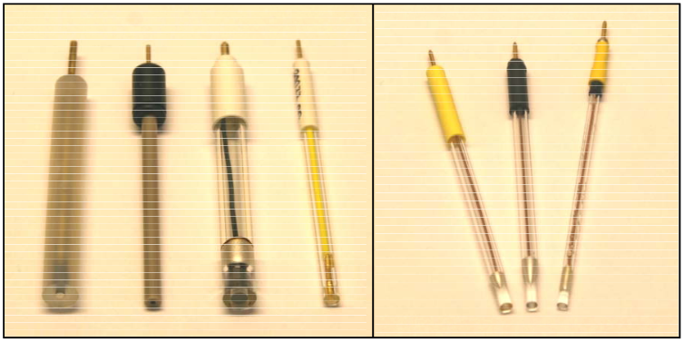
Figure 35
Electrodes with diameters less than about 25 μm, termed microelectrodes or ultramicroelectrodes, advanced by R. M. Wightman and coworkers offer unique electrochemical characteristics. These include, in addition to their extreme small size, minimization of solution resistance effects and rapid response times. Important applications of these electrodes are in very high speed voltammetry (>10,000 V/s), electrochemistry in highly resistive solvents, and in vivo voltammetry. Electrodes can be fabricated in the low μm diameter range by sealing platinum or gold microwires, or carbon fibers in glass. Electrodes at these dimensions can also be produced by metal deposition or photolithography.
The electrochemical behavior of microelectrodes can appear markedly different from that observed at conventionally sized electrodes. To illustrate the difference, we will first consider the case of a planar electrode of mm dimensions in a cell with a volume of several mL. If a change is made in the applied potential, either a step or a sweep, from a value where no electron transfer occurs to a redox active solution species, to one in which electron transfer occurs, the concentration of the redox active species will be lowered at the electrode surface, leading to the formation of a concentration gradient (see chronoamperometry, Section II A, part 1-a for review). The longer the electrode is at a potential sufficient to allow electron transfer, the further away from the electrode into solution the concentration gradient extends.
The existence of the gradient induces diffusion of electroactive material from regions of high concentration (the bulk of the solution) to regions of low concentration (near the electrode surface). This diffusion can be described by Fick’s Laws, which take slightly different forms for changing electrode geometries. For the large planar electrode described here, the diffusion layer moves very quickly far out into the solution, exceeding the distance a molecule can diffuse on the time scale of a typical experiment. Under these conditions, diffusion from the bulk of solution where concentration is constant to the electrode surface is nearly all linear in nature, in a direction perpendicular to the electrode surface.
In cyclic voltammetry (see Section II A, part 2-b) these conditions typically give rise to the traditional peak-shaped voltammogram. A CV recorded for ferrocene at a 3 mm diameter glassy carbon disk electrode is shown at the left side of Figure 36. Ferrocene was present at a concentration of 0.6 mM in acetonitrile with 0.1 M tetrabutylammonium hexafluorophosphate as supporting electrolyte. The scan rate was 0.10 V/s.
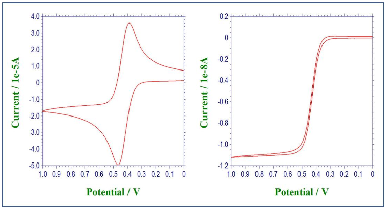
Figure 36
Next consider a planar microelectrode with micrometer or smaller dimensions. At the right side of Figure 36, a voltammogram for 0.6 mM ferrocene recorded at a glassy carbon electrode with a diameter of only 10 μm is shown. With all of the other experimental conditions remaining the same, a sigmoidal rather than a peak-shaped voltammogram was observed. This was the result of a steady-state condition between diffusion and electron transfer, where the rate of diffusion matches the rate of electron transfer. Why the difference? Because of the small size of the electrode, the contribution to the current by diffusion from the edges of the electrode becomes important in the total mass transport of electroactive species. This edge effect, or radial diffusion is generally very small at large electrodes relative to the linear diffusion mentioned above. For microelectrodes, the flux per unit time and area is greater than for large electrodes because the region from which electroactive species diffuses to the surface is in essence hemispherical in shape.
It is important to understand that the voltammograms in Figure 36 are for one set of conditions. There are conditions in which a CV recorded at a large planar electrode will demonstrate steady-state behavior, and conditions for which peak-shaped voltammograms are seen at microelectrodes. The normalizing factor for all conditions is the experimental time. The approximate distance (in cm) a diffusing molecule can travel during a time period t (in seconds) is given by
\[\mathrm{d = (2\, D_0\, t)^{1/2}}\]
where D0 is the diffusion coefficient (cm2/s). When d is small relative to the radius of the electrode, linear diffusion will predominate, and the observed voltammogram will be peak shaped. For small electrode dimensions, d will frequently be large relative to the electrode radius, and a steady-state voltammogram will result.
While this has been only a cursory look at the differences between macro- and micro-sized electrodes, there are many excellent review articles available for readers desiring more in-depth information on this topic.25-28
3. Electrode Preparation
Ideally, a working electrode should behave reproducibly each time that it is used. Factors that affect the electrochemical behavior of a surface are its cleanliness, the kind and extent of chemical functionalities (including oxides) that are present, and the microstructure of the electrode material itself. Generally, a pretreatment step or steps will be carried out prior to each experiment to assure that the surface going into the electrochemical cell can be reproduced experiment-to-experiment. These may be as simple as mechanical polishing, and may include pre-scanning across a certain potential range or exposure to a solvent or chemical species to “activate” the electrode. Specific procedures for different electrode materials can be found in References 29-30 and those contained therein.