
Quantum Mechanical Description of NMR Spectroscopy
- Page ID
- 79325

The first thing that must be considered is the nature of a hydrogen atom.
What makes up the nucleus of a hydrogen atom?
Hopefully you remember from general chemistry that the nucleus of a normal hydrogen atom consists of a single proton. Hydrogen has two other isotopes: (1) deuterium, which has one proton and one neutron in its nucleus, and (2) tritium, which has one proton and two neutrons in its nucleus. Our development will focus only on normal hydrogen with a single proton in its nucleus.
Thinking back to the coverage of the nature of electrons in atoms, you learned that electrons are described by a series of quantum numbers (principal, angular, magnetic, and spin) and that the electrons in atoms could be described using electronic configurations (1s2, 2s2, 2p6, etc.). It turns out that the particles in the nucleus of an atom are also described through a set of quantum numbers and that the protons and neutrons in a nucleus are described using a nuclear configuration. Understanding the exact form of a nuclear configuration is not important in understanding NMR spectroscopy (this knowledge would be of interest to a nuclear chemist studying nuclear decay processes such as alpha, beta and gamma decay). What is important is that nuclear particles spin like electrons spin and therefore nuclear particles have spin quantum numbers. The value I is used to denote the total spin quantum number for a nucleus.
What are the allowable spin quantum numbers for an electron?
Hopefully you remember that the two allowable spin quantum numbers are +½ and –½.
What do you think are the allowable spin quantum numbers for a proton?
Perhaps it will make intuitive sense that they will also be +½ and –½.
What is the magnitude of I, the total nuclear spin, for a hydrogen nucleus?
Since the hydrogen nucleus only has a single proton with a spin quantum number of ½, the value of I is also ½.
What do you think is produced by the spinning, charged proton that is the hydrogen nucleus?
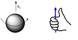
Any spinning charged object generates a magnetic dipole (i.e., magnetic field). Magnetic fields are designated by the symbol B, so we can designate the magnetic field produced by the hydrogen nucleus as Bp. Magnetic fields also have an orientation to them. The orientation is determined using what is known as the right-hand rule. The proton spins about an axis as shown on the left in Figure 1. To determine the orientation of the magnetic field, curl the fingers of your right hand in the direction of the spin and the right thumb points in the direction of the magnetic field (Figure 1). Magnetic fields are represented using vectors and the vector representation for Bp is also shown in Figure 1.
NMR samples are placed in a magnet and therefore subjected to an applied magnetic field (BAPPL). It is important to note that the magnitude of BAPPL is significantly larger than the magnitude of Bp.
What happens when two magnetic fields (Bp and BAPPL) are in contact with each other?
At some point in your life you have probably played with two magnets and know that they interact with each other. In one orientation, the two magnets attract. In another orientation, they repel each other. This means that the proton’s magnetic field must interact with BAPPL. This interaction constrains Bp to only certain allowable orientations relative to BAPPL. The number of allowable orientations of a nucleus in an applied magnetic field is (2I + 1). Since I = ½ for the hydrogen nucleus, there are two allowable orientations.
What do you think are the two allowable orientations of Bp relative to BAPPL?
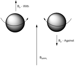
As shown in Figure 2, one orientation has Bp aligned “with” and the other has Bp aligned “against” the applied magnetic field. Note: the length of the vectors shown in Figure 2 is not an accurate representation of the actual magnitude of the two magnetic fields. BAPPL is so much larger than Bp that an accurate vector representing BAPPL would be so large it could not fit onto the page (alternatively, if the size of the vector for BAPPL was accurate, the vector for Bp would be so small it would not be visible). Also note from the pictures in Figure 2 that the two different orientations involve the proton spinning in opposite directions relative to BAPPL.
Do you think the two allowable orientations have the same or different energy?
Perhaps it intuitively makes sense that the two will have different energies. If the energies were not different, it would not be possible to record NMR spectra.
Which of the two do you think is lower in energy?
Again, it might seem intuitive that the orientation in which Bp is “with” BAPPL would be lower in energy, which turns out to be the case.
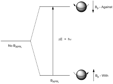
This now allows us to draw an energy level diagram as shown in Figure 3. In the absence of an applied magnetic field the hydrogen nucleus only has a single possible energy. In the presence of an applied magnetic field, the hydrogen nucleus has two allowable energy states: the lower energy one being the ground state, the higher energy one being the excited state. As with other energy level systems you have previously encountered, photons of electromagnetic radiation with an energy that exactly matches the energy difference between the ground and excited state have the ability to excite the nucleus from the ground spin state (Bp “with” BAPPL) to the excited spin state (Bp “against” BAPPL).
One thing worth considering is the exact nature of what happens to the proton as it is excited from the ground spin state to the excited spin state. Remember that the difference in the two states is that the proton spins in opposite directions. One possibility is that the proton literally stops spinning to reverse course and spin in the opposite direction. But another possibility is that the proton continues spinning but flips upside down relative to an external observer. To try this out, hold a ball in your hands and spin it about an axis in a particular direction, then flip it over while still maintaining the spin. You will note that, from your view, it now spins in the opposite direction. This spin flip is what actually happens to a hydrogen nucleus when it is excited from the ground to excited state.
We know from quantum mechanics that Eq 1 applies to such a system:
\[ΔE = hν \tag{1}\]
This means that a discrete frequency of electromagnetic radiation is needed to cause the excitation transition from the ground to the excited state (flip the spin of the proton).
What frequency of electromagnetic radiation is needed to excite a nuclear spin flip?
The energy gap shown in Figure 3 for nuclear spin flips corresponds with the radiofrequency portion of the electromagnetic spectrum. Note, radio frequencies occur in the megahertz (MHz) portion of the spectrum. Perhaps your school has an FM radio station. If so, its broadcast frequency will be in the MHz range. Also, your department probably has something like a 60, 400 or 600 MHz NMR spectrometer.
Where is radiofrequency (RF) radiation on the energy scale of the electromagnetic spectrum?
RF radiation is at the very low energy end of the spectrum of electromagnetic radiation. This exceptionally small gap in energy between the ground and excited state has several significant consequences in NMR spectroscopy that will be developed in this unit. In order to understand some of these consequences, we need to consider how the energy difference between the ground and excited nuclear spin states compared to the energy that is available to any chemical system at ambient (i.e., room) temperature. This is referred to as thermal energy and is denoted by kT.
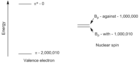
Consider the energy gap for a valance electron transition (e.g., \(\pi\)-\(\pi\)*) that occurs in the ultraviolet part of the electromagnetic spectrum as it compares to the energy gap for nuclear spin flips that occurs in the RF part of the spectrum. The energy levels in Figure 4 illustrate this difference, although the gap for the \(\pi\)-\(\pi\)* transition is actually shown much smaller than it should be so it fits onto the page.
Is the thermal energy at room temperature large or small compared to the energy of a \(\pi\)-\(\pi\)* transition and to the energy of a nuclear spin flip? What are the consequences of your answers to these questions?
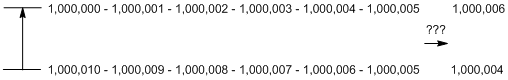
The thermal energy at room temperature is small compared to the energy of a \(\pi\)-\(\pi\)* transition but is large compared to the energy of a nuclear spin flip. This observation means that thermal energy is insufficient to excite a valence electron from its ground to excited state and cannot promote an electron from a \(\pi\) to a \(\pi\)* orbital. However, thermal energy is sufficient to excite nuclear spin flips. The consequence of this observation involves the populations of the ground and excited states for the two systems. For a \(\pi\)-\(\pi\)* system, all of the species will be in the ground state because thermal energy is insufficient to cause excitation. For a nuclear spin system, thermal energy is sufficient to cause the transition from the spin-with to spin-against state and cause population of the excited nuclear spin flip state. As an example, let’s consider the possible populations in a representative system of 2,000,010 chemical species. Populations of energy states can be calculated using the Boltzmann distribution. As shown in Figure 4, for the \(\pi\)-\(\pi\)* system, all 2,000,010 are in the ground state. For the nuclear spin flip system, 1,000,010 are shown in the ground state and 1,000,000 are in the excited state.
If thermal energy has sufficient energy to excite nuclear spin flips, why are there still more in the ground than excited state?
The reason there are still more in the ground state than the excited state is that it is lower in energy and chemical systems have a preference for lower energy states than higher energy states. But it is important to recognize that in NMR spectroscopy, the populations of the two energy states are about equal. This will have some important consequences. One relates to the sensitivity of NMR spectroscopy. Another relates to something known as coupling.
Suppose we now send in the exact frequency match to excite the nuclear spin flip illustrated in Figures 4 and 5. This will begin to excite nuclei up to the excited state, which will begin to change the populations as shown in Figure 5. Excited state nuclei have extra energy and want to decay or relax back to the ground state.
Can you think of two processes by which a specific excited state nucleus can get rid of its excess energy?
The first of these involves the loss of the energy to the surroundings as heat. This is known as spin-lattice or longitudinal relaxation and is denoted as T1. Spin-lattice relaxation reestablishes the original populations of the two levels.
To understand the second, we need to consider that the excited state nucleus has an amount of extra energy that is exactly what is needed to excite a ground state nucleus. A process known as spin-spin or transverse relaxation can occur where this energy is essentially transferred from an excited state nucleus to a ground state nucleus. The result is that the nucleus that started in the excited state ends up in the ground state and the nucleus that started in the ground state ends up in the excited state. Note that this mechanism does not reestablish the original population distribution.
Do excited state nuclei have short or long relaxation times?
Perhaps your intuition says that, because the energy gap between the ground and excited state is so small, a nucleus does not gain much by relaxing back to the ground state so that the relaxation time is relatively long. This reasoning is correct with the observation that the typical spin-lattice relaxation time for a 1H nucleus is on the order of 1-2 seconds.
A consequence of the long relaxation times is that, if you apply lots of photons with the frequency needed to excite the nuclei, they are excited up faster than they relax back down. The result is that the populations of the two levels quickly equalize as shown in Figure 5 where each has 1,000,005.
When the populations of the two levels are equal, can we continue to excite ground state nuclei up to the excited state such that the population of the excited state becomes larger than the population of the ground state, creating what is known as a population inversion?
Producing a population inversion is essential to the functioning of a laser, but a laser has more than the two levels (ground and single excited state) that occur with the nuclear spin flip in NMR. We need to examine this 2-level system in more detail.
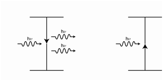
At the point where the populations are equal in Figure 5, incident photons have an equal probability of interacting with a ground state or excited state nucleus. A photon that interacts with the ground state is absorbed and excitation occurs. A photon that interacts with the excited state leads to something called stimulated emission. Stimulated emission occurs when an excited state system interacts with an incident photon and the energy of the photon exactly matches the energy gap between the excited and ground state. In the process of stimulated emission, the extra energy of the excited state system is emitted as a photon and the incident photon is also emitted – so two photons of exactly the same energy come out. Figure 6 shows the process involved when an incident photon interacts with the excited state resulting in stimulated emission (left-hand side) and when the incident photon interacts with the ground state resulting in absorption (right-hand side). The net result in Figure 6 is that, when the populations of the ground and excited states are equal, two photons come in and two come out. To an external observer it now appears that no photons are being absorbed. This resonance is said to be saturated.
A saturated resonance produces no signal because there is no net absorption of photons. One necessity to reduce the likelihood of saturating resonances is to use a low power for the RF source, which means fewer incident photons are put into the sample. Because it is so easy to saturate NMR resonances, the sensitivity of the method is much lower than other spectroscopic methods. This means that NMR measurements require more concentrated samples than other spectroscopic methods such as UV/Visible absorption spectroscopy. However, there are other factors that influence the sensitivity of NMR that are important to consider.
One important observation is that the difference in energy between the ground and excited states is proportional to the magnitude of the applied magnetic field, so that a larger applied magnetic field increases the difference in energy between the ground and excited state (Figure 7).
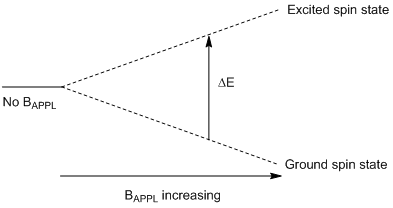
What happens to the population distribution as the energy gap between the ground and excited state is increased?
The population distribution is proportional to the size of the energy gap, such that using a larger applied field leads to a more favorable population distribution and more sensitivity. For example, if the magnetic field strength was high enough such that the population of the ground state was 1,000,040 and excited state 999,970, then 35 nuclei can be excited before the resonance is saturated. The enhanced sensitivity is one reason why many investigators want NMR spectrometers with a high magnetic field strength.
Another feature that can be used to enhance the sensitivity is to use microtubes (from 5 to 35 μl depending on the specific features of the instrument). The smaller volume of the microtube compared to a conventional NMR tube (about 600 μl) means that a smaller weight of sample is needed for solutions of the same concentration. This can be especially helpful for analyzing small amounts of a natural product isolated from a living organism. Another feature that enhances the sensitivity of NMR spectrometers is to use a cryoprobe. The probe is the component of the instrument containing the sample and the electronics for inputting the RF and measuring signal. “Cryo” refers to the use of very cold temperatures, either at liquid helium (4 K or −269°C) or liquid nitrogen (77 K or −195.79°C) temperature. The sample itself is not cooled but the electronic components used to transmit and receive the signal are cooled. Cooling these components leads to a significant reduction in the electronic noise thereby leading to a 2- to 5-fold enhancement in the signal-to-noise ratio (S/N). One final feature in NMR spectrometers that can be used to enhance the S/N is to record the spectrum multiple times and add these together. Since noise is random, some of the noise cancels out when several spectra are added together. Signal is additive, therefore the S/N increases.
We have already noted that ΔE = hν for the nuclear spin flip transition (Eq 1). Eq 2 shows the further relationship of the energy gap of the transition to other parameters in which γ is the magnetogyric ratio of the nucleus and Bo is the magnetic field experienced by the nucleus.
\[ΔE = \dfrac{hγ B_o}{2π} \tag{2}\]
The magnetogyric ratio is a fundamental parameter of a nucleus and is defined as shown in Eq 3, in which μ is the magnetic moment and I is the total spin quantum number for the nucleus.
\[γ= \dfrac{2πμ}{hI} \tag{3}\]
Substituting Eq 3 into Eq 2 and rearranging to solve for only the frequency of excitation gives Eq 4.
\[ν = \dfrac{μB_o}{hI} \tag{4}\]
For I = ½ nuclei such as hydrogen, Eq 5 is obtained for DE.
\[∆E=2μB_o \tag{5}\]
It is important to note that the magnetic moment is constant for a particular nucleus. In other words, all 1H nuclei, no matter what their surrounding environment, have exactly the same magnetic moment. All 13C nuclei, which also have a spin of ½, have exactly the same magnetic moment, but the magnetic moment of the 13C nucleus is only one-fourth as large as that of the 1H nucleus. Since Eq 4 and Eq 5 also apply to 13C nuclei, the difference in the magnetic moment between 1H and 13C means that the energy gap and resonant frequency of a 13C nucleus is one-fourth of the energy gap and resonant frequency of a 1H nucleus. NMR spectrometers are designated by the frequency (e.g., 400 MHz) needed to excite the spin flips of 1H nuclei rather than the size of BAPPL. Note, magnetic fields are in units of Tesla and the applied magnetic field that corresponds with a 400 MHz excitation frequency for the 1H nucleus is 9.4 Tesla. On a 400 MHz spectrometer, the excitation frequency of 13C nuclei is 100 MHz because its magnetic moment is one-fourth that of the 1H nucleus.
Since the magnetic moment is the same for all 1H nuclei, the only term that varies in Eq 4 and Eq 5 for a 1H nucleus is the Bo term. It is important to note that Bo is proportional to BAPPL but is not identical to BAPPL. Instead, Bo is the sum of all the magnetic fields that are experienced by a particular nucleus. One magnetic field to consider is that of the earth. The magnitude of the earth’s magnetic field may vary at different places but it exists and nuclei within an NMR spectrometer will experience it. So we could write Eq 6 as a first example that distinguishes Bo from BAPPL. However, since BEarth will be a constant at any particular location, this term would not traditionally be included in an equation representing Bo.
\[B_o= B_{APPL} + B_{Earth} \tag{6}\]
Consider a sample in an NMR tube. The crosshatched region in the tube in Figure 8 is the area over which signal is recorded. Why is it important that BAPPL be homogeneous over this entire region?
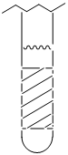
If we examine Eq 4 and Eq 5, the only variable is Bo, which is proportional to BAPPL. If BAPPL is different for different regions of the NMR tube, then Bo will be different for those regions. This means that the frequency needed to excite a specific hydrogen nucleus of a molecule varies over these regions. Differences in the frequency needed to excite a specific hydrogen nucleus means that the resonance will be broadened in the spectrum. Two steps are taken to reduce this broadening. One is to tune (also called shimming) the NMR spectrometer. Magnetic fields have a direction to them. When tuning, the X, Y and Z parameters are adjusted to optimize the homogeneity of BAPPL over the region of the NMR tube where signal is measured. The other step is to spin the sample. Spinning causes a specific hydrogen nucleus in a specific molecule to experience BAPPL over a range of sites as it spins through them. This averaging improves the homogeneity of BAPPL over the entire sample.
NMR spectra are usually recorded with deuterated (2H) solvents such as chloroform-d (CDCl3). One reason for replacing normal hydrogen with deuterium in the solvent is to reduce the size of the solvent signal in the spectrum. The other is that tuning is usually done on the deuterium signal. Deuterium is an NMR active nucleus, but its magnetic moment is different from 1H so it resonates at a different frequency on the spectrometer. This makes it possible to monitor the 2H signal to optimize the tuning while not perturbing the 1H spectrum that is being measured. The spectrometer is also locked onto the deuterium signal so, if any drift of the signals were to occur while the spectrum is being recorded, it will be accounted for.
What “things” in a molecule generate magnetic fields that will influence Bo for a particular hydrogen nucleus?
Earlier in this unit we determined that spinning charged particles generate a magnetic field. Electrons and protons in molecules are charged and have spin. Therefore, we might reason that electrons and protons in molecules may generate a magnetic field that influences the magnitude of Bo. Electrons in molecules produce an effect known as shielding. Protons in molecules produce an effect known as coupling. Shielding determines where resonances are located in the spectrum. Coupling determines the shape of the resonance. Without these differences in the location and shape of resonances, NMR spectra would be useless for determining the structures of molecules. We can now write Equation 7 for Bo in which Be represents the magnetic field produced by electrons and Bp represents the magnetic field produced by protons. However, we must consider the details of shielding and coupling to better understand the specific features of NMR spectra.
\[B_o= B_{APPL} + B_e + B_p \tag{7}\]