
10.3.6: Fluorescence Microscopy
- Page ID
- 257595
An important use of fluorescence spectroscopy techniques is in the area of microscopy, and this is especially true in the realm of biology and biochemistry. In this section a brief description of fluorescence microscopy will be presented with an emphasis on techniques to improve the resolution.
A fluorescence microscope is much the same as a conventional light microscope with added features to enhance its capabilities.
- The conventional microscope uses visible light (400-700 nanometers) to illuminate and produce a magnified image of a sample.
- A fluorescence microscope, on the other hand, uses a much higher intensity light source which excites a fluorescent species in a sample of interest. This fluorescent species in turn emits a lower energy light of a longer wavelength that produces the magnified image instead of the original light source.
Fluorescent microscopy is often used to image specific features of small specimens such as microbes. It is also used to visually enhance 3-D features at small scales. This can be accomplished by attaching fluorescent tags to anti-bodies that in turn attach to targeted features, or by staining in a less specific manner. When the reflected light and background fluorescence is filtered in this type of microscopy the targeted parts of a given sample can be imaged. This gives an investigator the ability to visualize desired organelles or unique surface features of a sample of interest.
A block diagram of a typical wide field fluorescence microscope and a picture of a readily available fluorescence microscope is shown in the Figure\(\PageIndex{1}\).
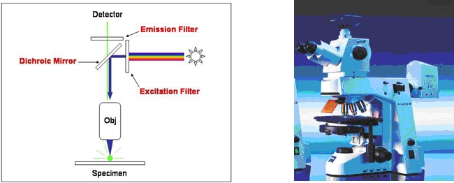
Figure\(\PageIndex{1}\): A schematic diagram of a wide-field fluorescence microscope and a picture of a typical commercially available fluorescence microscope. these images were taken from Fluorescent Microscopy by George Rice, Montana State University (https://serc.carleton.edu/microbelif.../fluromic.html)
A wide field microscope contains a detector capable of recording the entire image in a single collection (a digital camera) without moving the sample. A dichroic mirror is a mirror that transmits one wavelength (color) of light but reflects another wavelength (color). Most fluorescence microscope come equipped with a small set of dichroic mirrors appropriate for a small set of fluorescence labels. Like all simple optical microscopes the spatial resolution is limited to approximately \({\lambda}\). However, the emitted light is collected from regions above and below the focal plane of the microscope objective leading to a lesser ability to discern sharp features in the sample.
The first major improvement in terms of resolution for a fluorescence microscope was the invention of the confocal fluorescence microscope. As shown in the Figure\(\PageIndex{2}\), a pinhole has been introduced before the detector to spatially limit the volume from which the fluorescence is collected. The pinhole optical element along with the microscope objective give this instrument two focal points along the light path. Light is collected only from the focal point of the microscope objective and emitted light from above and below the focal point of the microscope objective is discriminated (blocked) by the focal point created by the pinhole. Images are collected by moving the sample in a point by point raster pattern and the image is reconstructed from the signal at each point. Confocal microscope are commercially available albeit at a much higher cost relative to a wide field fluorescence microscope.
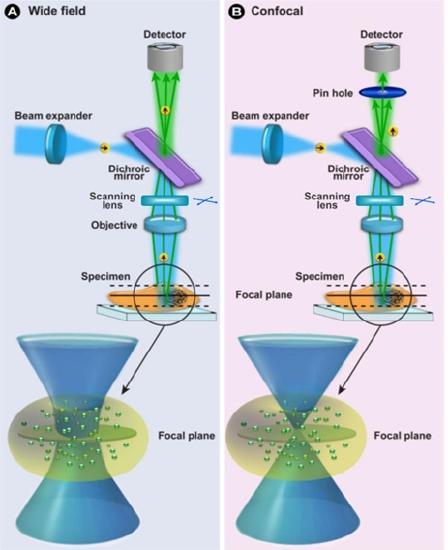
Figure\(\PageIndex{2}\): An illustration of a confocal fluorescence microscopy along side an illustration of a wide field fluorescene microscopy. This image was adapted from one found in New approaches in renal microscopy - Scientific Figure on ResearchGate. Available from: https://www.researchgate.net/figure/...fig2_299444264
Another major improvement in terms of resolution comes with the two-photon fluorescence microscope. A two photon fluorescence microscope employs a laser that emits light for which a single photon is insufficiently energetic enough to of excite the fluorophore. Because of the high spectral brightness of the laser, when the light focused by the microscope objective an intense electric field is created at the focal point and the energy associated with two photons is sufficient to excite the fluorophore. The difference between one photon excitation with short wavelength light and two photon excitation with long wavelength light is schematically shown in F Figure\(\PageIndex{3}\).
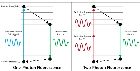
Figure\(\PageIndex{3}\): An energy level diagram illustrating the difference between one photon excitation with short wavelenght light and two photon excitation with long wavelength light.
Because the electric field is sufficiently large only at the focal point the emission from the excited fluorophore only comes from this small volume region. Figure\(\PageIndex{4}\) is a picture showing the fluorescence in a one and two photon experiment in a cuvette. In Figure 10.3.6.4 each absorbed photon at 488 nm is capable of exciting the fluorophore and hence the emission is observed coming from a very large volume region. The 960 nm is insufficient of exciting the fluorophore with a single photon and only excites the fluorophore in the small volume region at the focal point .
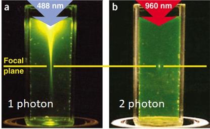
Figure\(\PageIndex{4}\): In this photo the small volume region in which the excited fluorophores are produced with two photon excitation with 960 nm light is compared to the large volume region in which the excited fluorophores are produced with one photon excitation with 488 nm light. This image was taken from Zipfel, W., Williams, R. & Webb, W. Nonlinear magic: multiphoton microscopy in the biosciences. Nat Biotechnol 21, 1369–1377 (2003). https://doi.org/10.1038/nbt899
A two photon fluorescence microscope is depicted in Figure\(\PageIndex{5}\) where red excitation beam shown is coming from a laser. Again in this type of fluorescence microscope the image is generate by translating the sample in a point by point raster pattern, collecting the signal at each point and reconstructing the image.
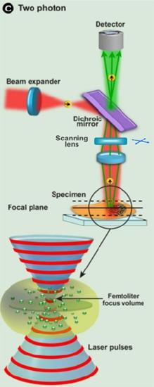
Figure\(\PageIndex{5}\): An illustration of a two photon fluorescenc microscope. This image was adapted from one found in New approaches in renal microscopy - Scientific Figure on ResearchGate. Available from: https://www.researchgate.net/figure/...fig2_299444264
Figure\(\PageIndex{6}\) shows a series of fluorescent images of a pollen sample illustrating the spatial resolution and ability to probe the sample as a function of depth for the three types of fluorescence microscopes described in this section. It should be readily observable that the confocal and two-photon microscopes provide far superior images to a standard wide field fluorescence microscope.
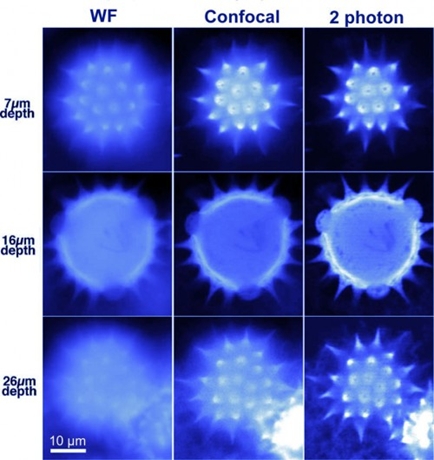
Figure\(\PageIndex{6}\): Fluorescent pollen grain was imaged at three depths below its surface (7, 16, and 26 μm) by wide field, confocal, and 2 photon technique. Two-photon imaging improves image sharpness for deep optical sections by reducing scattering of excitation light and by eliminating fluorescence excitation outside the plane of focus. This image was taken from http://candle.am/microscopy/.