
2.5: A Closer Look
- Page ID
- 211472
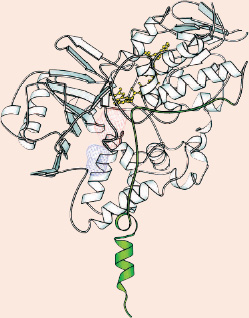
One protruding end (green) of the MAO B enzyme anchors the protein inside the cell. Body molecules or drugs first come into contact with MAO B (in the hatched blue region) and are worked on within the enzyme's "active site," a cavity nestled inside the protein (the hatched red region). To get its job done, MAO B uses a helper molecule (yellow), which fits right next to the active site where the reaction takes place.
REPRINTED WITH PERMISSION FROM J. BIOL. CHEM. (2002) 277:23973-6 HTTP://WWW.JBC.ORG
Seeing is believing. The cliché could not be more apt for biologists trying to understand how a complicated enzyme works. For decades, researchers have isolated and purified individual enzymes from cells, performing experiments with these proteins to find out how they do their job of speeding up chemical reactions. But to thoroughly understand a molecule's function, scientists have to take a very, very close look at how all the atoms fit together and enable the molecular "machine" to work properly.
Researchers called structural biologists are fanatical about such detail, because it can deliver valuable information for designing drugs—even for proteins that scientists have studied in the lab for a long time. For example, biologist have known for 40 years that an enzyme called monoamine oxidase B (MAO B) works in the brain to help recycle communication molecules called neurotransmitters. MAO B and its cousin MAO A work by removing molecular pieces from neurotransmitters, part of the process of inactivating them. Scientists have developed drugs to block the actions of MAO enzymes, and by doing so, help preserve the levels of neurotransmitters in people with such disorders as Parkinson's disease and depression.
However, MAO inhibitors have many undesirable side effects. Tremors, increased hart rate, and problems with sexual function are some of the mild side effects of AMO inhibitors, but more serious problems include seizures, large dips in blood pressure, and difficulty breathing. People taking MAO inhibitors cannot eat foods containing the substance tyramine, which is found in wine, cheese, dried fruits, and many other foods. Most of the side effects occur because drugs that attach to MAO enzymes do not have a perfect fit for either MAO A or MAO B.
Dale Edmondson of Emory University in Atlanta, Georgia, has recently uncovered new knowledge that may help researchers design better, more specific drugs to interfere with these critical brain enzymes. Edmonson and his coworkers Andrea Mattevi and Claudia Binda of the University of Pavia in Italy got a crystal-clear glimpse of MAO B by determining its three-dimensional structure. The researchers also saw how one MAO inhibitor, Eldepryl®, attaches to the MAO B enzyme, and the scientists predict that their results will help in the design of more specific drugs with fewer side effects.

Got It?
Define metabolism.
How does aspirin work?
Name three functions of blood.
Give two examples of immunotherapy.
What is a technique scientists use to study a protein's three-dimensional structure?